Download
1 / 4
50 likes | 95 Views
Metal oxide is highly important material which possesses many unique optical and electrical properties for applications in many areas such as Solar cells, Gas sensors and so on. With the development of research and applications of Metal oxide thin films, research results are verified that the morphology of Metal oxide thin films are plays an important role in applications of these films. Variety of morphologies, complex structure has been developed by physical or chemical methods. However the work on controlled growth of these films is still in developing state. Therefore in present work we deposited ZnS and ZnO metal oxides thin films on different substrates by Chemical Bath Deposition Technique. Structural, Surface Morphology and Optical properties of as deposited films were investigated by XRD, SEM, and UV VIS Spectrophotometer. The band gap is also calculated from the equation relating absorption co efficient to wavelength. The band gap indicates the film is transmitting within the visible range and the band gaps changes because of the grain size of the films. We also observed that, the change in preparative parameters affects the deposition rate of thin films. From the observation, it is clear that the growth rate increases as the deposition temperature, increases. S. S. Kawar "Preparation and Properties of Nanocrystalline Zinc Oxide Thin Films" Published in International Journal of Trend in Scientific Research and Development (ijtsrd), ISSN: 2456-6470, Volume-4 | Issue-4 , June 2020, URL: https://www.ijtsrd.com/papers/ijtsrd31623.pdf Paper Url :https://www.ijtsrd.com/physics/nanotechnology/31623/preparation-and-properties-of-nanocrystalline-zinc-oxide-thin-films/s-s-kawar<br>

E N D
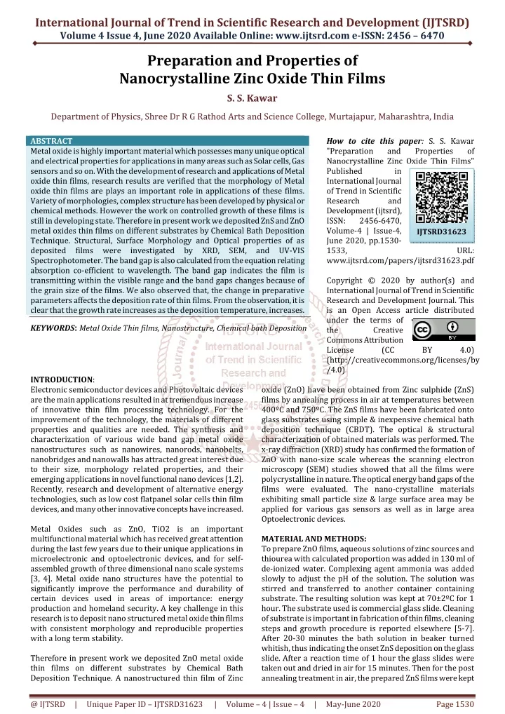
International Journal of Trend in Scientific Research and Development (IJTSRD) Volume 4 Issue 4, June 2020 Available Online: www.ijtsrd.com e-ISSN: 2456 – 6470 Preparation and Properties of Nanocrystalline Zinc Oxide Thin Films S. S. Kawar Department of Physics, Shree Dr R G Rathod Arts and Science College, Murtajapur, Maharashtra, India ABSTRACT Metal oxide is highly important material which possesses many unique optical and electrical properties for applications in many areas such as Solar cells, Gas sensors and so on. With the development of research and applications of Metal oxide thin films, research results are verified that the morphology of Metal oxide thin films are plays an important role in applications of these films. Variety of morphologies, complex structure has been developed by physical or chemical methods. However the work on controlled growth of these films is still in developing state. Therefore in present work we deposited ZnS and ZnO metal oxides thin films on different substrates by Chemical Bath Deposition Technique. Structural, Surface Morphology and Optical properties of as deposited films were investigated by XRD, SEM, and UV-VIS Spectrophotometer. The band gap is also calculated from the equation relating absorption co-efficient to wavelength. The band gap indicates the film is transmitting within the visible range and the band gaps changes because of the grain size of the films. We also observed that, the change in preparative parameters affects the deposition rate of thin films. From the observation, it is clear that the growth rate increases as the deposition temperature, increases. KEYWORDS: Metal Oxide Thin films, Nanostructure, Chemical bath Deposition How to cite this paper: S. S. Kawar "Preparation and Nanocrystalline Zinc Oxide Thin Films" Published in International Journal of Trend in Scientific Research and Development (ijtsrd), ISSN: 2456-6470, Volume-4 | Issue-4, June 2020, pp.1530- 1533, www.ijtsrd.com/papers/ijtsrd31623.pdf Copyright © 2020 by author(s) and International Journal of Trend in Scientific Research and Development Journal. This is an Open Access article distributed under the terms of the Creative Commons Attribution License (CC (http://creativecommons.org/licenses/by /4.0) Properties of IJTSRD31623 URL: BY 4.0) INTRODUCTION: Electronic semiconductor devices and Photovoltaic devices are the main applications resulted in at tremendous increase of innovative thin film processing technology. For the improvement of the technology, the materials of different properties and qualities are needed. The synthesis and characterization of various wide band gap metal oxide nanostructures such as nanowires, nanorods, nanobelts, nanobridges and nanowalls has attracted great interest due to their size, morphology related properties, and their emerging applications in novel functional nano devices [1,2]. Recently, research and development of alternative energy technologies, such as low cost flatpanel solar cells thin film devices, and many other innovative concepts have increased. Metal Oxides such as ZnO, TiO2 is an important multifunctional material which has received great attention during the last few years due to their unique applications in microelectronic and optoelectronic devices, and for self- assembled growth of three dimensional nano scale systems [3, 4]. Metal oxide nano structures have the potential to significantly improve the performance and durability of certain devices used in areas of importance: energy production and homeland security. A key challenge in this research is to deposit nano structured metal oxide thin films with consistent morphology and reproducible properties with a long term stability. Therefore in present work we deposited ZnO metal oxide thin films on different substrates by Chemical Bath Deposition Technique. A nanostructured thin film of Zinc oxide (ZnO) have been obtained from Zinc sulphide (ZnS) films by annealing process in air at temperatures between 400ºC and 750ºC. The ZnS films have been fabricated onto glass substrates using simple & inexpensive chemical bath deposition technique (CBDT). The optical & structural characterization of obtained materials was performed. The x-ray diffraction (XRD) study has confirmed the formation of ZnO with nano-size scale whereas the scanning electron microscopy (SEM) studies showed that all the films were polycrystalline in nature. The optical energy band gaps of the films were evaluated. The nano-crystalline materials exhibiting small particle size & large surface area may be applied for various gas sensors as well as in large area Optoelectronic devices. MATERIAL AND METHODS: To prepare ZnO films, aqueous solutions of zinc sources and thiourea with calculated proportion was added in 130 ml of de-ionized water. Complexing agent ammonia was added slowly to adjust the pH of the solution. The solution was stirred and transferred to another container containing substrate. The resulting solution was kept at 70±2ºC for 1 hour. The substrate used is commercial glass slide. Cleaning of substrate is important in fabrication of thin films, cleaning steps and growth procedure is reported elsewhere [5-7]. After 20-30 minutes the bath solution in beaker turned whitish, thus indicating the onset ZnS deposition on the glass slide. After a reaction time of 1 hour the glass slides were taken out and dried in air for 15 minutes. Then for the post annealing treatment in air, the prepared ZnS films were kept @ IJTSRD | Unique Paper ID – IJTSRD31623 | Volume – 4 | Issue – 4 | May-June 2020 Page 1530
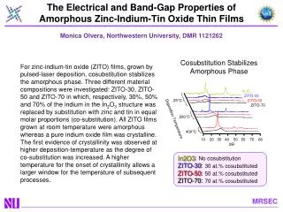
International Journal of Trend in Scientific Research and Development (IJTSRD) @ www.ijtsrd.com eISSN: 2456-6470 in the oven at various temperatures between 400ºC and 750ºC for 10 hours. The ZnS films get oxidized in the oven to form thin films of ZnO. The crystallographic structure of films was analyzed with x- ray diffractometer (EXPERT-PRO) by using Cu-Kα lines (λ= 1.542Å). The average grain size in the fabricated films was obtained from a Debye-Scherrer’s formula. Surface morphology was examined by JEOL model JSM-6400 scanning electron microscope (SEM). The optical transmission spectra for a range of samples were obtained in UV-VIS-NIR region using Perklin-Elmer UV-VIS lambda-35 spectrometer.[8-9]. RESULTS AND DISCUSSION: Structural properties: Figure1. shows the X-ray diffraction pattern of ZnS thin films and ZnO thin film obtained from post annealing treatment over ZnS films at various temperatures. The XRD pattern confirms that the ZnS films get oxidized in the oven to form ZnO thin films. It was found that cubic structured ZnS is completely oxidized at higher annealing temperature to form a hexagonal ZnO. The film shows reflections from (100), (002) and (101) planes at 600°C, indicating the formation of ZnO nano particles having pure hexagonal structure (matches with JCPDF data) [9]. 800 Meas. data:S1 Meas. data:ZnS Sample1 600 500 600 In ten s ity (c p s) In ten sity (c ps ) 400 400 300 200 200 100 0 0 100 100 In ten s ity (cp s ) In ten s ity (c p s) 50 0 0 -50 -100 -100 20 40 60 80 20 40 60 80 2-theta (deg) 2-theta (deg) Figure1. XRD patterns of ZnS and ZnO thin film fabricated at 600ºC The average size of grain (g) have been obtained from the XRD patterns using Debye-Scherrer’s formula, [9] g = Kλ / β cosθ ------------- (1) Where, K = constant taken to be 0.94, λ = wavelength of X-ray used (1.542Å), β = FWHM of the peak and θ = Bragg’s angle. The grain sizes were found to be within the range from 08 to 43nm. This confirms the good crystallinity of the samples. Determination of Grain size and lattice strain of ZnS thin films are shown in Table1. S. No. Sample 2 Theta (Degree) FWHM Value (Degree) Grain size (nm) Lattice strain 1 ZnS 1 59.89 1.17 32.62 32.295 37.077 48.263 57.31 63.55 68.56 0.47 Table1: Determination of grain size & lattice strain of thin film sample Surface Properties: The surface morphology of deposited ZnO thin films were investigated by SEM at different magnifications as shown in Figure 2. 8.19 39.31 43.43 32.97 33.69 25.56 37.089 27.36 0.0089 0.0033 0.0030 0.0040 0.0026 0.0030 0.0015 0.0030 0.22 0.199 0.262 0.27 0.37 0.30 2 ZnS 2 Figure 2 Scanning Electron Micrograph of as deposited ZnO thin film at different magnification. The deposits are compact, pinhole free with spherical grains from few nanometers up to clusters of 157 nm and the films are well covered on the substrate. From the figure, it is observed that the small nanosized grains engaged in a fibrous- like structure and nanoribbons which clearly indicates the nanocrystalline nature along with some amorphous phase of ZnO thin films. The average grain size of the nanoparticles is about 130 nm. [10-11]. @ IJTSRD | Unique Paper ID – IJTSRD31623 | Volume – 4 | Issue – 4 | May-June 2020 Page 1531
International Journal of Trend in Scientific Research and Development (IJTSRD) @ www.ijtsrd.com eISSN: 2456-6470 Optical Properties: The optical energy band gap (Eg) was determined by plotting (αhν)2 versus hν and then extrapolating the straight line portion to the energy axis at α =0 (Figure 3). It was observed that the band gap of the film obtained at higher annealing temperature is 2.31eV [12-14]. Figure3. Plot of (αhν)2 vs hν for ZnO thin films fabricated at 600ºC Effect of Deposition Temperature on Film Thickness: Figure 4 shows the variation of film thickness as a function of deposition time at different temperatures. Various temperatures from 3130K to 3450K in steps of 100K were used in the depositing ZnS & ZnO to obtain the optimal temperature. Figure 4 shows an increase in film thickness as the temperature increases from 3130K to 3450K. The rise in the film thickness may be due to the dissociation of the Zinc-complexes (with ammonia and with thiourea) and increase in the hydrolysis of SC(NH2)2 as the temperature increases. At higher temperatures, the decomposition of thiourea will be faster setting more S2- ions free. Additionally, the Zinc-complex dissociation is greater and gives higher concentrations of free Zn2+ in the solution, which in turn results in higher deposition rates or in other words for a constant buffer layer thickness, in shorter deposition times. Also the kinetic energy of the ions in solution is higher at higher temperature, which brings about increased interaction between them and subsequent deposition at volume nucleation centers of the substrate. It can be seen from figure, the average growth rate is high at 345ºK (9.478nm/min) and it is slow at 313ºK (7.622nm/min). The prepared ZnS films were kept in the oven at various temperatures between 400ºC and 750ºC for 10 hours. The ZnS films get oxidized in the oven to form thin films of ZnO [15]. 40C 60C 70C 90C x=0.15 PH=10 R.P.M.=60/MIN R.T.=35C 280 260 300 240 220 250 200 thickness(nm) 180 thickness(nm) 200 160 140 150 120 100 100 80 50 60 10 20 30 40 50 60 70 80 90 100 30 40 50 60 70 80 90 time(min) deposition temperature C FIGURE 4: Variation of film thickness as a function of deposition time at different temperatures. CONCLUSION: In present work we deposited ZnS and ZnO metal oxides thin films on different substrates by Chemical Bath Deposition Technique. Structural, Surface Morphology and Optical properties of as deposited films were investigated by XRD, SEM, and UV-VIS Spectrophotometer. The X-ray diffraction analysis showed that film is polycrystalline with pure hexagonal structure. The grain size estimated is in the range of 08 to 43nm. The band gap is also calculated from the equation relating absorption co-efficient to wavelength. The band gap indicates the film is transmitting within the visible range and the band gaps changes because of the grain size of the films. The film has a direct band gap with an optical value of 2.31eV which is in good agreement with the standard value. We also observed that, the change in preparative parameters affects the deposition rate of thin films. From the observation, it is clear that the growth rate increases as the deposition temperature increases. This makes the films suitable for various optoelectronic and sensor applications. References: [1]J. Y. Lao, J. Y. Huang, D. Z. Wang, Z. F. Ren, D. Steeves, B. Kimball, and W. Poter, Appl. Phys. A Materials Science& Processing, 78, 539, 2004; [2]O. K. Varghese and C. A. Grimes, Journal of Nanoscience and Nanotechnology, 3, 277, 2003. [3]H. J. Fan, W. Lee, R. Scholz, A. Dadgar, A. Krost, K.Nielsch,and M. Zacharias, Nanotechnology, 16,913, 2005. @ IJTSRD | Unique Paper ID – IJTSRD31623 | Volume – 4 | Issue – 4 | May-June 2020 Page 1532
International Journal of Trend in Scientific Research and Development (IJTSRD) @ www.ijtsrd.com eISSN: 2456-6470 [4]Y. Xia, P. Yang, Y. Sun, Y. Wu, B. Mayers, B. Gates, Y. Yin, F. Kim, and H. Yan, Adv. Mater. 15, 353, 2003. [11]G. K. Williamson, W. H. Hall, Acta Metall. Vol. 1, PP. 22– 31, 1953. [5]P. A. Radi, A. G. Brito, J. M. Madurro, and N. O. Dantas Brazilian J. of Physics, Vol.36, PP.412-414, 2006. [12]J. J. Kelly, D. Vanmaekelbergh, in: G. Hodes (Ed.), Electrochemistry of Nanomaterials, Wiley- VCH, Weinheim, pp. 103–136, 2001. [6]J. P. Borah and K. C. Sarma, Acta Physica Polonica A Vol. 114, No.4, 2008. [13]G. Cao, Nanostructure and Nanomaterials, first ed., Imperial College Press, London, 2004. [7]M. Y. Nadeem, Waqas Ahme, Turk J Phy Vol. 24, PP. 651,2000. [14]P. X. Gao and Z. L. Wang, Journal of Applied Physics, 97,044304, 2005. [8]Jie Cheng, DongBo Fan, et al, Semicond. Sci. Technol. Vol.18, PP. 676, 2003. [15]K. K. Hurde, A. B. Lad, S. S. Kawar, B. H. Pawar, International Journal of Materials Science, Volume 12, Number 1, pp. 9-16 , 2017. [9]S. S. Kawar, B. H. Pawar, Chalcogenide Letters, Vol. 6, No. 5, p. 219 – 225, 2009 [10]B. S. Remadevi, R. Raveendran, A. V. Vaidyan, Prammana J. Phy., Vol. 68, PP. 679,2007. @ IJTSRD | Unique Paper ID – IJTSRD31623 | Volume – 4 | Issue – 4 | May-June 2020 Page 1533