Download

1 / 20
1.14k likes | 3.82k Views
Retinal Disorders. By: Nassar Nassar. ANATOMY. Retina as seen through an ophthalmoscope. Gross Anatomy. Nervous coat of the eyeball Outer surface is attached to the choroid Inner surface is attached to the vitreous body Ends as wavy line – ora serrata Macula:

E N D
Retinal Disorders By: Nassar Nassar
Gross Anatomy • Nervous coat of the eyeball • Outer surface is attached to the choroid • Inner surface is attached to the vitreous body • Ends as wavy line – oraserrata • Macula: • Oval, yellowish area at center of posterior part • High concentration of cones • It includes the Fovea Centralis • Optic Disk: • 3mm medial to macula • Pierced by the central artery of retina • Blind Spot • Leave as optic nerve
Layers • Outermost: retinal pigment epithelium (RPE) – single layer and is not part of the retina • Most books say htat the retina is 10 layers but grossly it is infact 3 layers. • Innermost: neuroretina: • Ganglion cell layer, axons form optic nerve • Bipolar nerve layer • Photoreceptors (cons & rods)
Photoreceptors • Convert light into electrical signals • Rods: • For night vision • Do not signal wavelength information (color) • Cones: • For daylight and color vision • High threshold to light • Concentrated at the fovea
Direction of Light Light pass through the ganglion and bipolar layers before reaching the photoreceptors in all areas of the retina except the fovea. In the fovea, the bipolar and ganglion cell layers are pulled aside so that light strikes the photoreceptors directly. Visual Processing
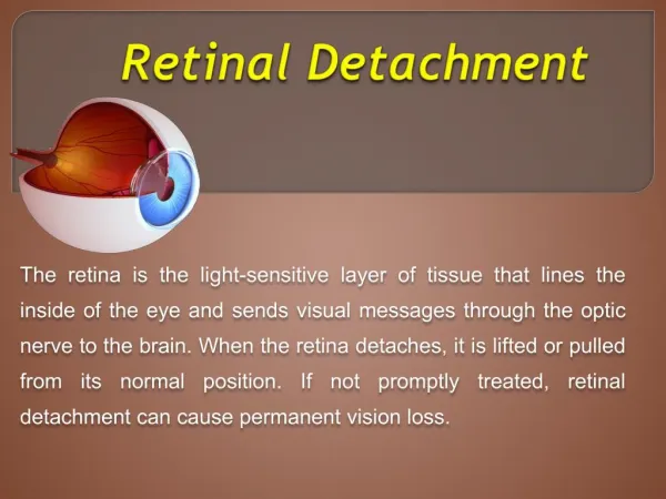
Macular Dysfunction Disorder of the central part of the macula (fovea) causes significant visual impairment. The patient may complain of: • Blurred central vision • Distorted vision (metamorphopsia): micropsia or macropsia, occur if the photoreceptors become stretched apart or close together. • Areas of loss of central visual field (scotomata), if part of the photoreceptor layer becomes covered, e.g. by blood, or if the photoreceptors are destroyed.
Blurred central vision Scotomata Metamorphopsia
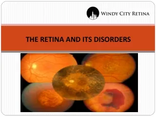
Peripheral Retinal Dysfunction The pt. complains of: • Loss of visual field, detected clinically. • Some diseases may predominantly affect one type of photoreceptors, ex. Retinitis pigmentosa and night vision.
Age-related macular degeneration (AMD) • The commonest cause of irreversible visual loss in the developed world. • Associated with increasing age and is typically bilateral • Pathogenesis: • Overtime undigested lipid products deposits in Bruch’s membrane, seen as yellow lesions called drusen. • Collection of drusen in the macula is termed age related maculopathy (ARM) • Dry form: the neighboring RPE and photoreceptors show degenerative changes • Wet/Exudative form: angiogenic factors (ex. VEGF) stimulate new vessel formation from the choroid through Bruch’s membrane and RPE into the sub retinal space forming sub-retinal neovascular membrane.
AMD cont. Symptoms • Symptoms of macular dysfunction • Progressive, gradual loss of central vision leading to difficulty in reading and recognizing distant objects • In the wet form, visual disturbance is sudden Signs • Yellow, well-circumscribed drusen • Areas of hypo/hyperpigmentation • Loss of foveal reflex • In wet, pre-retinal (more occasionally) or subretinal hemorrhage
AMD cont. Investigations • Diagnosis is based on the appearance of the retina. • In suspected exudative AMD and vision not severely affected a fluorescein angiogram may be performed to delineate the position of the sub-retinal neovascular membrane. Prognosis • Dry AMD: progress very slowly , increasing difficulty in reading • Wet AMD: 75% of pt.s experience marked deterioration of vision over a 3 year time frame.
AMD cont. Wet AMD Fluorescein Angiogram of Wet AMD