Download
1 / 36
390 likes | 681 Views
Ovaries & Testes, Sperm & Egg (and their reproduction), and Fertilization/Early Development. Kayla Lovett, Katie Mackin , Alex Brockdorf. Reproduction . Ovary and Testes. Function of various testis cells.

E N D
Ovaries & Testes, Sperm & Egg (and their reproduction), and Fertilization/Early Development Kayla Lovett, Katie Mackin, Alex Brockdorf
Reproduction Ovary and Testes
Function of various testis cells • Interstitial cells (Leydig cells): secrete testosterone and other androgens (type of steroid that includes testosterone) in the presence of LH. • Germinal epithelium cells: stratified cellular cover. Line seminiferous tubules, epididymis, vans deferens, ejaculatory duct, bulbourethral glands, seminal vesicle. • Developing spermatozoa: maturing spermatids that will become spermatozoan (basically baby sperm that are growing) • Sertoli cells: long, striated cells. Provide support, protection, and nutrition for developing spermatids, which detach when mature. Activated by FSH. IB Standard 11.4.1
Function of various ovarian cells • Germinal epitheliuim: Layer of epithelial cells lining the ovaries. Basically same in males, but for ovaries. • Primary follicles: Contain primary oocyte. Basically just primary oocyte plus some epithelial cells. Changes to secondary oocyte thanks to LH and FSH. • Mature follicle • Secondary: contains secondary oocyte and follicular fluid • Vesicular (Graafian): still secondary oocyte. Fluid filled cavity grows to the point that follicle walls balloon to surface of ovary. Bursts and releases secondary oocyte (ovulation). Forms corpus luteum after ovulation. • Secondary oocyte: Commonly known as egg. Located in mature follicles. Where the sperm penetrates to become a full egg. IB Standard 11.4.4
Reproduction Sperm and Egg
Sperm IB Standard 11.4.6
Sperm Anatomy • 1. Tail: flagellum, which allows sperm to swim toward egg • 2. Middle Piece: contains mitochondria-produces energy • 3. Head: contains nucleus, and a membrane-bound acrosome, which contains enzymes needed to penetrate the egg IB Standard 11.4.6
Egg IB Standard 11.4.6
Egg Anatomy • Corona radiata: first layer of follicular cells, just outside of zonapellucida • Zonapellucida: extracellular matrix, just outside of the plasma membrane. IB Standard 11.4.6
Reproduction Oogenesis and Spermatogenesis
Oogenesis • Defined as the production of eggs in females by the process of meiosis and maturation. • Occurs in the ovaries • Starts after an ovarian stem cell (oogonia) undergoes mitosis to form a primary oocyte w/46 chromosomes. • Two major stages: Meiosis I and Meiosis II • Occur after mitosis and a period of cellular growth IB Standard 11.4.5
Meiosis I • Primary oocytes with 46 chromosomes • Two haploid cells with 23 chromosomes are produced • One of these haploid cells is a Secondary Oocyte (egg) • Bigger because it gets most of the cytoplasm • The other haploid is a Polar Body • Essentially ways to dispose of chromosomes while egg retains cytoplasm • Could either disintegrate or divide again IB Standard 11.4.5
Meiosis II • Secondary Oocyte (egg) goes through Meiosis II but stops at metaphase. • Takes a journey from the ovary to the oviduct where one of two things could happen: • Sperm could be present- meiosis II completed and another polar body forms (fertilization) • No sperm present- disintegrate without completing meiosis II • Fertilization in this stage restores diploid number of chromosomes- fusion of egg and sperm nuclei. IB Standard 11.4.5
Spermatogenesis • Defined as production of sperm in males by the process of meiosis and maturation. • Occurs in the seminiferous tubules in the testes after stem cells (spermatogonia) supply primary spermatocytes. • Three stages: Meiosis I, Meiosis II, and Metamorphosis and maturation • Occur after mitosis and a period of cell growth IB Standard 11.4.2
Meiosis I • Starts with primary spermatocytes with 46 chromosomes • Two secondary spermatocytes form, each with 23 duplicated chromosomes IB Standard 11.4.2
Meiosis II • Starts with secondary spermatocytes • Produces four spermatids with 23 daughter chromosomes IB Standard 11.4.2
Metamorphosis and Maturation • Spermatids differentiate into actual sperm • Then, upon sexual arousal, those sperm exit upon ejaculation. IB Standard 11.4.2
LH, Testosterone, FSH in Spermatogenesis • FSH: follicle stimulating hormone • Promotes spermatogenesis • LH: luteinizing hormone • Controls production of androgen testosterone by interstitial cells (between seminiferous tubules) • Involved in negative feedback relationship that maintains fairly constant testosterone/sperm production • Testosterone: main male sex hormone • Necessary for maturation of sperm IB Standard 11.4.3
Spermatogenesis vs. Oogenesis • Number of gametes: millions-produced all throughout lifetime. • Timing of Formation: Continuous • Timing of Release: Anytime • Begins/Ends: at puberty/through whole life (slows down) • Meiotic divisions uninterrupted • Number of gametes: fixed-only about 400 mature • Timing of Formation: once a month (menstrual cycle) • Timing of Gamete Release: monthly cycle • Begins/Ends: during fetal development/menopause • Arrested meiotic divisions IB Standard 11.4.8
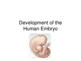
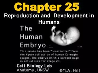
Reproduction Fertilization & Early Development (up to implantation of blastocyst)
Fertilization • 1. Arrival of sperm: attracted by a chemical signal, swim up oviduct to reach egg • 2. Binding: first sperm to break through follicle cells binds to zonapellucida • 3. Acrosome reaction: acrosomal cap separates from sperm and contents are released. Proteases digest route through zonapellucida which allows sperm to reach plasma membrane of the egg • 4. Fusion: plasma membranes of sperm and egg fuse and sperm nucleus enters egg and joins egg nucleus • 5. Corticol reaction: cortical granules move to plasma membrane and fuse with it, releasing contents by exocytosis. Enzymes from cortical granules cause cross linking of glycoproteins in zonapellucida, hardening it to prevent entry of more sperm • 6. Mitosis: nuclei of sperm and egg carry out mitosis, producing a two cell embryo IB Standard 11.4.9
Embryonic Development: The First Week • Fertilization occurs in the upper oviduct • Cleavage: begins 30hrs after fertilization, continues as embryo goes through oviduct to uterus. • Morula: what embryo is when it reaches uterus on third day. Not much larger than zygote because of lack of cell growth. • Blastocyst: morula transforms by about fifth day. Has a fluid filled cavity, a single layer of outer cells, and inner cell mass. • Trophoblast: single layer of outer cells. Nourishes embryo. Later, reinforced by mesoderm layer, gives rise to chorion(extraembryonic membrane) • Inner cell mass eventually becomes embryo, then fetus
The Second Week • Implantation: trophoblast secretes enzymes to digest some of the tissue/blood vessels of endometrium. Embryo about size of a period. • HCG: hormone secreted by trophoblast. Basis for pregnancy test, maintains corpus luteum past time it normally disintegrates. Therefore maintains endometrium and prevents menstruation from occurring. • Formation of extraembryonic membranes • Yolk sac: forms below embryonic disk. No nutritive function, but is first site of blood cell formation. • Amnion: where embryo and fetus develop. Amniotic fluid insulates and absorbs shock. • Gastrulation • Embryonic disk: flattened inner cell mass. 2 layers of cells- ectoderm and endoderm. • Mesoderm: third layer of embryonic disk cells, forms once disk elongates into primitive streak. • Chorion: mesoderm reinforces trophoblast, which transforms into this. • These germ layers enable development of future organs
The Third Week • Nervous system development • Neural folds • Neural tube: later develops into brain and nerve cord. Formed when neural folds meet at midline. • Nerve chord: called spinal chord after notochord is replaced by vertebral column • Development of the heart: begins in third week and goes onto fourth. Right and left heart tubes fuse and begin to pump blood. Veins enter posteriorly, and arteries exit anteriorly. Later twists so all vessels are anteriorly.
The Fourth and Fifth Weeks • Mesoderm: forms a bridge called the body stalk which connects the tail with the chorion • Caudal: tail of embryo • Chorion: extraembryonic membrane formed by mesoderm-reinforced trophoblast • Chorionic villi: fingerlike projections which eventually form placental sinus. Contains allantois membrane, whose blood vessels become umbilical blood vessels • Umbilical cord formation: connects developing embryo to placenta • Limb buds: develop arms and legs, hands and feet • Head enlarges & sense organs become more prominent
The Sixth through Eighth Weeks • Becomes recognizable as human • Head develop normal relationship with body, neck region forms • Reflexes develop • 38 mm long, weighs as much as an aspirin tablet
Sources • http://s681.photobucket.com/user/akucic_biology/media/lolzballs.jpg.html • http://biology-10.wikispaces.com/file/view/sperm_cell.jpg/357526930/sperm_cell.jpg • http://images.tutorvista.com/content/reproduction-in-animals/human--ovary-top-section.jpeg • http://www.ib.bioninja.com.au/_Media/egg_and_sperm_med.jpeg • http://legacy.owensboro.kctcs.edu/gcaplan/anat2/notes/Image613.gif • http://www.bio.davidson.edu/Courses/Molbio/MolStudents/spring2005/Champaloux/fertilization.jpg