Download
1 / 1
10 likes | 76 Views
DIFFERENCES IN C-FOS EXPRESSION IN BASOLATERAL AND MEDIAL AMYGDALA IN CD-1 MICE FOLLOWING LPS INJECTION Research Project by Lianna Hrycyk, Supervised by Nafissa Ismail, Ph.D. Department of Behavioral Neurosciences, School of Psychology, University of Ottawa. METHODS. INTRODUCTION. CONCLUSION.

E N D
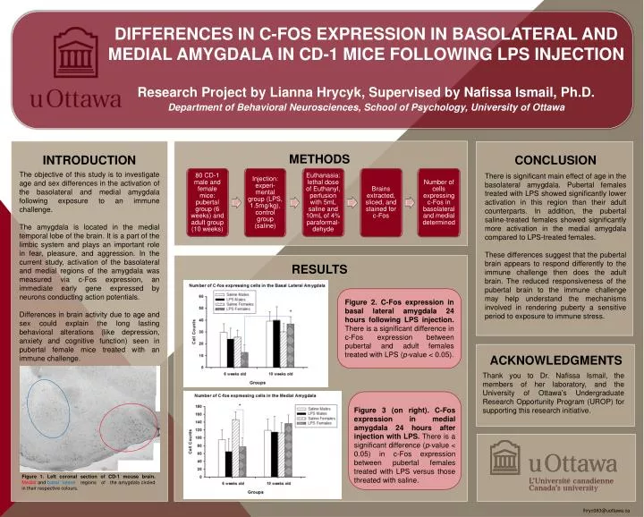
DIFFERENCES IN C-FOS EXPRESSION IN BASOLATERAL AND MEDIAL AMYGDALA IN CD-1 MICE FOLLOWING LPS INJECTION Research Project by Lianna Hrycyk, Supervised by Nafissa Ismail, Ph.D. Department of Behavioral Neurosciences, School of Psychology, University of Ottawa METHODS INTRODUCTION CONCLUSION The objective of this study is to investigate age and sex differences in the activation of the basolateral and medial amygdala following exposure to an immune challenge. The amygdala is located in the medial temporal lobe of the brain. It is a part of the limbic system and plays an important role in fear, pleasure, and aggression. In the current study, activation of the basolateraland medial regions of the amygdala was measured via c-Fos expression, an immediate early gene expressed by neurons conducting action potentials. Differences in brain activity due to age and sex could explain the long lasting behavioral alterations (like depression, anxiety and cognitive function) seen in pubertal female mice treated with an immune challenge. There is significant main effect of age in the basolateral amygdala. Pubertal females treated with LPS showed significantly lower activation in this region than their adult counterparts. In addition, the pubertal saline-treated females showed significantly more activation in the medial amygdala compared to LPS-treated females. These differences suggest that the pubertal brain appears to respond differently to the immune challenge then does the adult brain. The reduced responsiveness of the pubertal brain to the immune challenge may help understand the mechanisms involved in rendering puberty a sensitive period to exposure to immune stress. RESULTS Figure 2. C-Fos expression in basal lateral amygdala 24 hours following LPS injection. There is a significant difference in c-Fos expression between pubertal and adult females treated with LPS (p-value < 0.05). ACKNOWLEDGMENTS Thank you to Dr. Nafissa Ismail, the members of her laboratory, and the University of Ottawa’s Undergraduate Research Opportunity Program (UROP) for supporting this research initiative. Figure 3 (on right). C-Fos expression in medial amygdala 24 hours after injection with LPS. There is a significant difference (p-value < 0.05) in c-Fos expression between pubertal females treated with LPS versus those threated with saline. Figure 3 (on right). C-Fos expression in medial amygdala 24 hours after injection with LPS. There is a significant difference (p-value < 0.05) in c-Fos expression between pubertal females treated with LPS versus those threated with saline. Figure 1. Left coronal section of CD-1 mouse brain. Medial and basal lateral regions of the amygdala circled in their respective colours. lhryc083@uottawa.ca