Download
1 / 49
970 likes | 2.84k Views
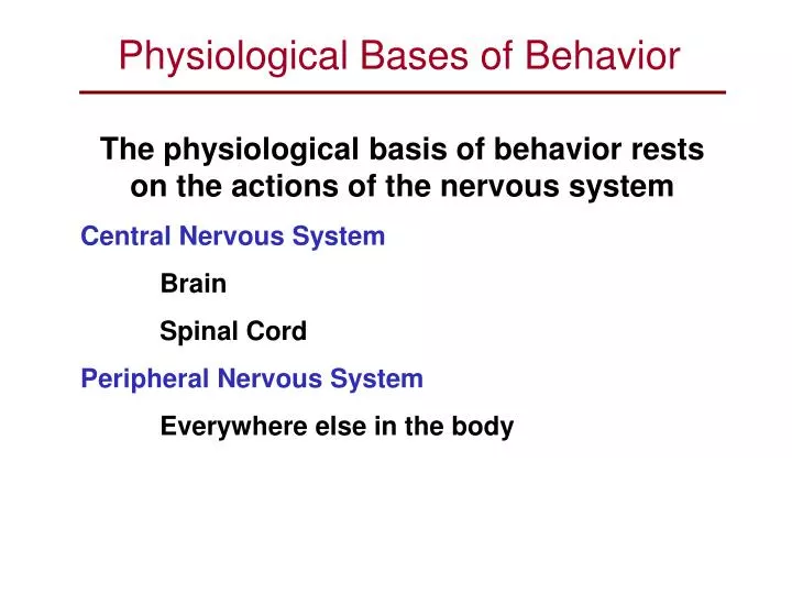
Physiological Bases of Behavior. The physiological basis of behavior rests on the actions of the nervous system Central Nervous System Brain Spinal Cord Peripheral Nervous System Everywhere else in the body. Electrical Activity of the Neuron. Cells of the Nervous System.

E N D
Physiological Bases of Behavior The physiological basis of behavior rests on the actions of the nervous system Central Nervous System Brain Spinal Cord Peripheral Nervous System Everywhere else in the body Electrical Activity of the Neuron
Cells of the Nervous System Receptor Cells or Sensory Cells (Afferent) Stimulated by events in our environment (Exteroceptors) or events that occur internally (Interoceptors) Interconnector Cells Provide pathways of communication and processing of information Response or Motor Cells (Efferent) Connected to muscles causing them to contract or relax thus controlling action Electrical Activity of the Neuron
The Reflex Arc The Bell-Magendie Law - Sensory nerves enter the spinal column from the dorsal side while motor nerves exit from the ventral side
The Neuron The cells of the nervous system Great variety of shapes and sizes Probably 100,000,000,000 in the human body Little replication capability in adults (Don’t Grow Back)
The Structures of the Neuron Soma (Cell Body) contains the Nucleus which is responsible for carrying out the metabolic activity of the cell thus producing the energy for its activity Dendrites are the receiving end of the neuron. At the dendrite the neuron receives stimulation from as many as 100 or so other neurons. Usually there are many dendrites Axon is the transmitting end of the neuron. Each neuron has only one axon although the end is branched. Axon diameter varies with thicker axons being capable of greater velocity of transmission (about 100m/sec) and thinner fibers being considerably slower (perhaps 1m/sec) Axon length varies tremendously with the longest axons in the human body being 3 ft or more in length (even longer in some big animals) A myelin sheath (fatty covering) surrounds most (but not all) axons. Supports the very long but very thin (fractions of a millimeter in diameter) axon. Insulates the axon electrically preventing cross-communication between pathways
Electrical Activity of the Neuron Information travels through the neuron in the form of an electrical impulse The electrical activity of the neuron follows through a number of sequential stages. In the Resting State there are, both inside and outside the neuron, fluids containing positively charged charged ions of both Sodium (Na+) and Potassium (K+). The concentration of positively charged ions is greater in the fluids outside the cell and the neuron is referred to as being polarized. The effects of stimulation on the resting neuron follow the All-Or-None Law (a neuron fires at full strength or not at all). There is no graduation of the electrical activity of the neuron. A neural impulse always occurs at full strength. Stimulation above the Excitatory Threshold causes an increase in permeability to Na+ The Action Potential refers to the depolarization (reversal of the usual electrical charge) that occurs when + charged sodium flows through the membrane wall into the interior of the neuron. The Action Potential causes depolarization of each successive segment of the axon as the neural impulse is propagated down the length of the axon. An active mechanism in the neuron called the Sodium- Potassium Pump returns the neuron back to normal by pushing Na+ and K+ out of the neuron. As the Sodium-Potassium Pump works to restore the resting state, the neuron passes through two consecutive refractory periods. The Absolute Refractory Period is a very brief period (1 msec) during which the neuron cannot produce another action potential regardless of the strength of stimulation applied. The Relative Refractory Period immediately follows the absolute refractory period. During this period another action potential can occur but a stronger than normally required (more intense) stimulus will be needed. May last as long as 8 msec. The Absolute Refractory period imposes a maximum firing rate for the neuron of about 1000 impulses per second. A very intense stimulus might cause firing at this maximum rate. Depending on stimulus intensity, another Action Potential might occur early or later in the Relative Refractory Period. Thus the neuron would respond to differences in the intensity of stimulation by variations in firing rate.
Chemical Activity of the Nervous System Information travels through the neuron in the form of an electric impulse but travels between neurons chemically. • The Synapse is the point of functional contact between neurons or between a neuron and a muscle • May involve thousands of neurons • Presynaptic Neuron contains the neurotransmitter in its axon terminal button (released into the Synaptic Clef during an Action Potential) • Postsynaptic Neuron is sensitive to and potentially stimulated by the neurotransmitter (excitatory) or inhibited by it (inhibitory) • Synaptic Transmission affected by neurotransmitters released into the synapse, chemicals resident in the synapse, and foreign chemicals from outside the body (Drug Effects)
Some of the Most Common Neurotransmitters Acetylcholine Norepinephrine Eprinephrin (Adrenalin) Serotonin Dopamine Probably two dozen others
Psychopharmacology All psychoactive drugs work by altering the normal processes at the synapse
Psychopharmacology • All psychoactive drugs work by altering the normal processes at the synapse • Drug Effects on Synaptic Transmission • Mimic the effects of a natural neurotransmitter • Prevent the uptake and synthesis of transmitters of their precursors • Influence (block or stimulate) the neurotransmitter release process or reuptake process • Block postsynaptic reception • Block neurotransmitter-degradative enzymes
The Brain • composed of two principal categories of cells: - neurons (100 billion) - neuroglia or glial cells (10 trillion) support and separate nerves cells as well as insulate nerve cell axons (myelin sheath). Also carry out phagocytosis (carry away dead cells and other debris)
Autonomic Nervous System (ANS) Responsible for maintenance of the internal environment (temperature, blood pressure, heart rate, hormone levels) Sympathetic Division – Its job is to mobilize the body’s resources for response to emergency. (Fight or Flight). Pupils Dilate, Respiration Increases, Heart Accelerates, Blood Pressure Rises, Adrenalin Secreted, Temperature Rises, Blood Flow shifted to muscles, piloerection, sweating, salivary and digestive shutdown. Parasympathetic Division – Its job is to replenish bodily resources (Relaxation). Pupils Constrict, Respiration Decreases, Heart Slows, Blood Pressure Falls, Temperature Falls, Blood Flow shifted to viscera, salivation returns to normal.
Comparative Neural Anatomy • Brain Size • Brain Size is directly proportional to the size of the cranium • Human brain = 1400cc Gorilla = 500cc Dolphin = 1600cc • Elephant = 5200cc • Degree of Convolution • The cortex of many animal species is relatively smooth. The cortex of more evolved animals is convoluted. • Differential Development of Structures • Species specific enhancement of different brain features (Sensory vs Cerebral)
Methods of Studying Brain Functioning • Lesion Techniques • Destruction, removal, or isolation of selected centers or structures of the brain. • Logic is destroy the area and see which behaviors disappear or become modified. Area destroyed must have been responsible for controlling the missing or altered behavior. • Electrolytic Lesion • Radio Frequency Lesion • Knife Cuts (Ablation) • Aspiration • Cryogenic Lesion
Methods of Studying Brain Functioning • Stimulation Techniques • Artificial activation of selected centers or structures of the brain. • Since the brain works both electrically and chemically, stimulation can be either electrical or chemical • Logic is activate the area and see which behaviors become modified. Area stimulated must have been responsible for controlling the elicited behavior.
The Stereotaxis • Stereotaxis used to stabilize subject's head while the procedure is being performed • Allows precise positioningof an electrode in a specific area of the brain • Stereotactic Coordinates from from an appropriate Brain Atlas
Histology Animal is Sacrificed Brain Removed From the Skull Brain Tissue Frozen Solid Microtome Slices Thin Cross-Sections of Tissue Tissue is mounted on Slides, Stained to reveal detail, and examined microscopically
Recording of Electrical Activity of the Nervous System • Can be used to study electrical activity of the brain is called an electroencephalograph (EEG) • Can be used to study tension of muscle group as an electromyograph (EMG) • Can be used to study heart functioning as an electrocardiograph (EKG) • Can be used to study movement patterns of the eyes as an electrooculagraph (EOG) • Can be used to study electrodermal activity or GSR (Galvanic Skin Response) Measures electrical conductivity of the skin. • Can also be used to study respiratory activity (Pneumograph)
The Homeostatic Drives Hunger Thirst Thermoregulation Excretion Respiration Sleep
Sleep and Wakefulness The body goes through a number of recurrent stages of alertness both during wakefulness and during sleep. Many of these stages recur about once per day (Circadian). Many patterns are shorter in a recurrence called the “Basic Rest and Activity Cycle” (BRAC) which is approximately 90-min in length.
Sleep Studies Electrodes are attached above an easily accessible muscle group to monitor the subject’s muscle tonus (EMG)
Stages of Sleep Slow Wave Sleep (NREM) has four stages which differ in depth and in EEG pattern
Stages of Sleep Stage 1 Sleep – lightest sleep, AAT (auditory awakening threshold) is very low, myoclonia occurs
Stages of Sleep Stage 2 Sleep – somewhat deeper, AAT is higher
Stages of Sleep Stage 3 Sleep – deep sleep, AAT is higher, difficult to wake subject from this stage
Stages of Sleep Stage 4 Sleep – very deep sleep, AAT is higher, very difficult to wake subject from this stage, capacity for stage 4 diminishes with age
Stages of Sleep Paradoxical Sleep (REM) – EEG suggests wakefulness but AAT is high, strong correlation with dream reports
REM Deprivation Studies Subjects wired with electrodes and monitored during an evening’s sleep. They were allowed to sleep as long as they wished except if the brain wave patterns showed REM sleep they were immediately awakened. This treatment continued for five consecutive evenings.
Effects of REM Deprivation After 5-Day REM Deprivation (Recover Period) Normal Cycle
REM Deprivation Studies Subjects wired with electrodes and monitored during an evening’s sleep. They were allowed to sleep as long as they wished except if the brain wave patterns showed REM sleep they were immediately awakened. This treatment continued for five consecutive evenings. Much longer REM deprivation studies have been conducted with animals (e.g., the cat)
REM Deprivation Studies Subjects wired with electrodes and monitored during an evening’s sleep. They were allowed to sleep as long as they wished except if the brain wave patterns showed REM sleep they were immediately awakened. This treatment continued for five consecutive evenings. Much longer REM deprivation studies have been conducted with animals (e.g., the cat) 175-day REM deprivation resulted in extreme REM rebound effects (100% above normal) but little else evident effect.
Insomnia – chronic problems getting adequate sleep Narcolepsy – sudden onsets of sleep during waking hours Sleep Apnea – reflexive gasping for air that awakens Somnambulism – sleepwalking Sleep Disorders