Download
1 / 49
500 likes | 524 Views
SHREE KRISHNA G N. INFECTIVE ENDOCARDITIS IN CHILDREN. Outline. Introduction Common cardiac conditions Causative organism Pathogenesis Clinical features Diagnosis, lab tests Treatment Prophylaxis. Objective of the lecture.

E N D
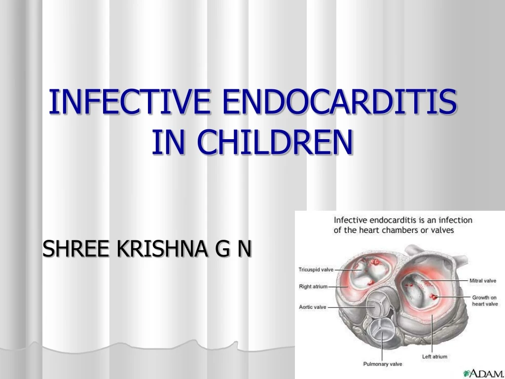
SHREE KRISHNA G N INFECTIVE ENDOCARDITIS IN CHILDREN
Outline • Introduction • Common cardiac conditions • Causative organism • Pathogenesis • Clinical features • Diagnosis, lab tests • Treatment • Prophylaxis
Objective of the lecture • At the end of the lecture a student should be able to write the following: • 1. pathogenesis of infective endocarditis • 2. clinical features of infective endocarditis • 3. diagnostic criteria • 4. management of infective endocarditis • 5. classify heart disease into high risk, • moderate and low risk groups to decide • prophylaxis.
DEFINITION Infection of the endocardial lining of heart. Endocardium of the valves Mural endocardium Endothelium of blood vessels Most common site is a diseased valve from where infection can spread to mural endocardium or vascular endothelium. IE occurs less commonly in children than in adults.
Common cardiac conditions- Congenital heart disease – VSD,PDA, aortic valve abnormalities, TOF, mitral valve prolapse. Previous corrective or palliative surgery for CHD with or without implanted vascular grafts patches or prosthetic cardiac valves. Rheumatic heart disease
Causative organisms could be bacteria, fungi or rickettsiae or viral. IE changes the prognosis of an otherwise benign lesion.
Risk factors 8-10% pediatric cases have no structural cardiac lesion or identifiable risk factor and involves mitral or aortic valve secondary to staph aureus. After dental procedure - Viridans group streptococci Lower bowel or genitourinary manipulations – group D streptococci (enterococci) IV drug abusers – pseudomonas aeroginosa, seracia marcescens Open heart surgery – fungal organisms No increased risk in immunocompromised child.
Organisms in pediatric IE Common native valve or other cardiac lesions Viridans group streptococci ( s. mutans, s. sanguis, s. mitis) Staphylococcus aureus Group D streptococcus ( Enterococcus).
Uncommon native valve or other cardiac lesions Streptococcus pnemoniae Hemophilus influenzae Coagulase negative staphylococci Coxiella bernetii ( Q fever) * N. gonorrhoea Brucella* Clamydia organisms* Legionella* Bartonella* HACEK group+ Sreptobacillus moniliformis* Pastorella multocida* Campylobacter fetus Culture negative ( 6% cases) * These are fastidius organism. These and fungi may produce culture negative endocarditis, special media, incubation for 7 days or serologic tests. + HACEK group ( H paraphrophilus, H. parainfluenza, H aphrophilus, Actinobacillus, Cardiobacterium hominis, Eikenella species, Kingella kingae)
Prosthetic valve Staphylococcus epidermidis Staphylococcus aureus Viridans group streptococci Pseudomonas aerogenosa Serracia mercescens Diphtheroids Legionella species HACEK group Fungi
IE in newborns Very high mortality rate. Incidence increased due to invasive techniques to manage newborns. Central lines and umbilical cannulation – right side of heart affected. Staph aureus, coagulase negative staph, candida, (rarely group B strep, S pneumoniae)
Pathogenesis Vegetations are formed at the site of endocardial or intimal erosion. Damaged endothelium is a potent inducer of thrombogenesis and provides a nidus to which bacteria can adhere and form vegetation. Combination of endothelial damage and bacteremia, development of IE.
PATOGENESIS POSTULATED BY WEINSTEIN AND SCHLESINGER NORMAL VALVE ENDOTHELIAL DAMAGE PLATELET DEPOSITION NONBACTERIAL THROMBOTIC ENDOCARDITIS (NBTE) BACTEREMIA COLONISATION OF NBTE INFECTIVE ENDOCARDITIS
Bacteria are trapped in the vegetations and are also in a quiescent stage, thus makes the organism less susceptible to antibiotics.
Clinical presentation Acute – usually staph aureus Sub acute – Viridans group streptococci ( alpha hemolytic), HACEK group ( H paraphrophilus, Actinobacillus, Cardiobacterium hominis, Eikenella species, Kingella kingae)
HISTORY Prior congenital or rheumatic heart disease Preceding dental, urinary tract or intestinal procedure Intravenous drug use Central venous catheter Prosthetic heart valve
SYMPTOMS Fever Chills Chest and abdominal pain Arthralgia, myalgia Dyspnoea Malaise Night sweats Weight loss CNS manifestations ( stroke, seizures, head ache) Usually insidious onset of illness, rarely fulminant and acute
Clinical findings relate to 4 underlying phenomena: Bacteremia or fungemia Valvulitis Immunologic responses Emboli
Extracardiac manifestations are relatively less common in children than in adults, eg : petichae, hemorrhages, Roth’s spots, Janeway lesions, Osler nodes, splenomegaly Renal abnormalities eg: glomerulonephritis, infarcts can result from embolic or immune complex mediated process.
Emboli to abdominal viscera, brain or heart may produce symptoms associated with ischemia, hemorrhage or both Uncommon: CNS mycotic aneurisms, their rupture can be catastrophic.
SIGNS Elevated temperature Tachycardia Roth’s spots, splinter nail bed hemorrhages, Osler nodes, Janeway lesions, petichae New or changing murmurs Splenomegaly Arthritis Heart failure Arrhythmias Metastatic infections ( arthritis, meningitis, mycotic arterial aneurism, pericarditis, abscesses, septic pulmonary emboli) Clubbing
Classic skin lesions Osler nodes –tender pea sized intradermal nodules in the pads of fingers and toes. Janeway lesions – painless small erythematous or hemorrhagic lesions on the palms and soles. Splinter hemorrhages – linear lesions beneath the nails. These lesion are seen late in the course and seldom seen in appropriately treated patients. These lesions represent vasculitis produced by circulating antigen antibody complexes.
Retinal hemorrhages: Roth’s spot – white or yellow center surrounded by a bright red irregular halo.
Janeway lesions Roth’s spot Osler nodes
Splinter hemorrhages Clubbing
Vegetation on tricuspid valve by echocardiography. Arrow denotes the vegetation.
Surgical excision of the vegetations on the tricuspid valve.
DUKES CRITERIA Major criteria – Positive blood culture for infective endocarditis Evidence of endocardial involvement
2. Evidence of endocardial involvement: Positive echocardiogram for IE Oscillating intracardiac mass on valve or supporting structures, or in the path of regurgitant jet, or on implanted material, in the absence of an alternative anatomic explanation or Abscess or New partial dehiscence of prosthetic valve, or new valvular regurgitation ( increase or change in pre-existing murmer not sufficient)
Minor criteria: Predisposing conditions ( heart disease, IV drug use) Fever >/= 38 degree C Vascular phenomena, a) major arterial emboli b) septic pulmonary infarcts c) mycotic aneurisms d) intracranial hemorrhage e) conjunctival hemorrhage f) Janeway lesions 4. Immunologic phenomena: a) glomerulonephritis b) Osler nodes c) Roth’s spot d) rheumatoid factor 5. Microbiological evidence: positive blood culture but not meeting major criteria 6. Serologic evidence of active infection with organisms consistent with IE 7. Echocardiogram: consistent with IE but not meeting major criteria.
Serologic tests: Rheumatoid factor ( > 50% positive in suacute endocarditis) Special serologic tests for Bartonella, Coxiella Polyclonal increase in gamma globulins characteristic of acute endocarditis Decreased complement in glomerulonephritis
2 major criteria or 1 major and 3 minor criteria or 5 minor criteria suggest definite endocarditis.
Laboratory tests Hemoglobin – subacute infection, normochromic normocytic anemia TC/DC – acute endocarditis, high granulocyte count with increase in band forms. ESR – elevated in 90%, may be low with heart or renal failure. CRP- usually elevated, falls to normal with treatment, better index than ESR Immune complexes Hypergammaglobulenemia Hypocomplementemia Cryoglobulenemia Renal failure – azotemia, high creat, Urinalysis – Microscopic hematuria and/ or slight proteinuria even without specific complicatins RBC casts and heavy proteinuria if immune complex GN occurs Gross hematuria in renal infarction
Blood cultures Most important test Should be obtained promptly 3-5 separate blood collections should be obtained Timing of collection not important as bacteremia is relatively constant In 90% cases causative organism is obtained from the 1st two cultures. Inform lab that IE is suspected so that culture is done on enriched media for longer than 7 days to detect nutritionally deficient and fastidious bacteria or fungi.
Chest Xray – bilateral infiltrates, nodules, pleural effusions. Echocardiography – evidence of valve vegetations, prosthetic valve dysfunction or leak, myocardial abscess, new onset valve insufficiency 2D ECHO Combination of transthoracic and transesophegeal ECHO
TREATMENT Can be considered under 2 headings: Treatment of current episode Prevention of endocarditis.
Principles of management are: Identification of organism Determining its antibiotic sensitivity Starting treatment as early as possible Using bactericidal antimicrobial agents for an appropriate duration to obtain cure and prevent relapse.
Fungal IE is treated with either amphotericin alone or its combination with flucystocine Surgical excision of the infected valve may be necessary. Relapses of endocarditis may occur.
PROPHYLAXIS Prophylaxis recommended: High risk- Prosthetic cardiac valves Previous bacterial endocarditis Complex cyanotic CHD Surgically constructed systemic pulmonary shunts or conduits Moderate risk- Most CHD other than those in high risk and negligible Acquired valvular dysfunction eg: rheumatic Hypertrophic cardiomyopathy MVP with valvular regurgitation and/or thickened leaflets
Prophylaxis not recommended: Negligible risk: ( same risk as general population) Isolated secundum ASD Surgical repair of ASD, VSD, PDA without residual defects and after 6 months of surgery Previous coronary artery bypass graft surgery MVP without valvular regurgitation Physiological, functional or innocent murmurs Previous Kawasaki disease without valvular dysfunction Previous rheumatic fever without valvular dysfunction Cardiac pacemakers and implanted defibrillators.
Further reading 1. Nelson, Textbook of Pediatrics, 18th edition 2. Essential Pediatrics, O.P.Ghai, 7th edition.
QUESTIONS 1)Name common organisms causing infective endocarditis?Write the clinical manifestation? 2)Enumerate DUKES CRITERIA? 3)Write about Treatment and Prophylaxis of infective endocarditis?
MCQ 1)Criteria used to diagnose infective endocarditis is? a)Jones criteria b) Nadas criteria c)Dukes criteria d) None. 2)Commonest cause of Endocarditis in iv drug user is ? a)Pseudomonas b)H Influenza. c)Brucella d)Legionella.
3)All are seen in infective endocarditis except? a)Erythema marginatum b)Oslers node. c)Janeway lesion d)Splinter hemorrage.