Download
1 / 22
220 likes | 287 Views
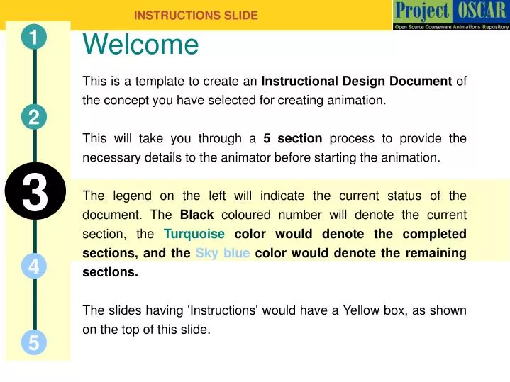
INSTRUCTIONS SLIDE. This is a template to create an Instructional Design Document of the concept you have selected for creating animation. This will take you through a 5 section process to provide the necessary details to the animator before starting the animation.

E N D
INSTRUCTIONS SLIDE This is a template to create an Instructional Design Document of the concept you have selected for creating animation. This will take you through a 5 section process to provide the necessary details to the animator before starting the animation. The legend on the left will indicate the current status of the document. The Black coloured number will denote the current section, the Turquoise color would denote the completed sections, and the Sky blue color would denote the remaining sections. The slides having 'Instructions' would have a Yellow box, as shown on the top of this slide. Welcome 1 2 3 4 5
Miss Hradhaya C Bhatlawande Protein Synthesis
Definitions and Keywords 1 • Translation-Translation is the production of proteins by decoding mRNA produced in transcription • Ribosome-complexes of RNA and protein that are found in all cells with nuclei. • transfer RNA (tRNA)-is a small RNA molecule (usually about 74-95 nucleotides) that transfers a specific active amino acid to a growing polypeptide chain at the ribosomal site of protein synthesis during translation • Codon-The genetic code is the set of rules by which information encoded in genetic material (DNA or RNA sequences) is translated into proteins (amino acid sequences) by living cells • Anticodon-base pair to the corresponding three base codon region on mRNA. 2 3 4 5
Shine-Dalgarno sequence-is a ribosomal binding site in the mRNA, generally located 16 nucleotides upstream of the start codon AUG. • wobble hypothesis-a wobble base pair is a non-Watson-Crick base pairing between two nucleotides in RNA molecules. • initiation factor-Initiation factors are proteins that bind to the small subunit of the ribosome during the initiation of translation, a part of protein biosynthesis • elongation factor-Elongation factors are a set of proteins that facilitate the events of translational elongation, the steps in protein synthesis from the formation of the first peptide bond to the formation of the last one • elongation factor Tu (EF-Tu) • elongation factor Ts (EF-Ts) • elongation factor G (EF-G) • molecular mimicry-Molecular mimicry is defined as the theoretical possibility that sequence similarities between foreign and self-peptides are sufficient to result in the cross-activation of autoreactive T or B cells by pathogen-derived peptides • release factor-The release factor is a protein that recognizes the termination codon or stop codon in a mRNA sequence on the ribosome
Translation-An Introduction • Translation is the whole process by which the base sequence of an mRNA is used to order and to join the amino acids in a protein. • The three types of RNA participate in this essential protein-synthesizing pathway in all cells; in fact, the development of the three distinct functions of RNA was probably the molecular key to the origin of life.
Translation An introduction(contd) • Three kinds of RNA molecules perform different but cooperative functions in protein synthesis • 1. Messenger RNA (mRNA) carries the genetic information copied from DNA in the form of a series of three-base code “words,” each of which specifies a particular amino acid. • 2. Transfer RNA (tRNA) is the key to deciphering the code words in mRNA. Each type of amino acid has its own type of tRNA, which binds it and carries it to the growing end of a polypeptide chain if the next code word on mRNA calls for it. The correct tRNA with its attached amino acid is selected at each step because each specific tRNA molecule contains a three-base sequence that can base-pair with its complementary code word in the mRNA • 3. Ribosomal RNA (rRNA) associates with a set of proteins to form ribosomes. • These complex structures, which physically move along an mRNA molecule, catalyze the assembly of amino acids into protein chains. • They also bind tRNAs and various accessory molecules necessary for protein synthesis. Ribosomes are composed of a large and small subunit, each of which contains its own rRNA molecule or molecules
Text for Step no. 1 • Initiation of protein synthesis in eukaryotic cells, as in bacteria, begins with formation of a preinitiation complex prior to mRNA binding. • Cells can regulate protein synthesis by phosphorylating a serine residue on the eIF2 bound to GDP; this complex is then unable to bind Met-tRNAiMet, thus inhibiting protein synthesis • The A preceding the AUG seems to be the most important nucleotide affecting initiation efficiency. • Scanning of the mRNA by the preinitiation complex eventually yields a initiation complex in which Met-tRNAiMet is correctly positioned at the translation start site.
Step no. 2 • The correctly positioned bacterial 70S ribosome Met-tRNAiMet complex is now ready to begin the task of stepwise addition of amino acids by the in-frame translation of the mRNA.
Text for Step No 2 • Once the small ribosomal subunit with its bound Met-tRNAiMet is correctly positioned at the start codon, union with the large ribosomal subunit completes formation of the initiation complex . • Initiation of translation of most mRNAs by theprokaryotic protein-synthesizing machinery begins near the 5′ capped end as just described.
Step No 3 • With the initiating Met-tRNAiMet at the P site and the second aminoacyl-tRNA tightly bound at the A site, the α amino group of the second amino acid reacts with the “activated” (aminoacylated) methionine on the initiator tRNA, forming a peptide bond
Step No 4 • For example, the second aminoacyl-tRNA is brought into the ribosome as a ternary complex in association with an EF-Tu – GTP in bacteria and becomes bound to the A site on the ribosome.
Step No 5 • the initiating Met-tRNAiMet is bound at the P site and base-paired with the AUG start codon. If the anticodon of the incoming (second) aminoacyl-tRNA correctly matches the second codon of the mRNA, a tight binding ensues at the A site. • If this second codon does not match the incoming aminoacyl-tRNA, it diffuses away. The choice of the correct aminoacyl-tRNA and its tight binding at the A site requires energy that is supplied by hydrolysis of the EF-Tu – GTP complex
Step No 6 • The key steps in elongation are entry of each succeeding aminoacyl-tRNA, formation of a peptide bond, and the movement, or translocation, of the ribosome with respect to the mRNA
During the process of peptide synthesis and tRNA site changes, the ribosome is moved along the mRNA a distance equal to one codon with the addition of each amino acid. This translocation step is catalyzed by bacterial EF-G – GTP, which is hydrolyzed to provide the required energy. Referring again to , we can see that after peptide linkage tRNAiMet, now without its activated methionine, is moved to an exit (E) site on the ribosome and is soon discharged. Concurrently, another ternary complex, carrying the next amino acid to be added, enters the ribosome, and the cycle continues.Protein Synthesis Is Terminated by Release Factors When a Stop Codon Is Reached RRF is another protein that resembles tRNA. The α helices of this protein mimic the tRNA structure. In contrast, in EF-G, β strands are the mimics, revealing an independent evolutionary origin. Step No 7
Step No 8 • The final phase of protein synthesis, like initiation and elongation, requires highly specific molecular signals that decide the fate of the mRNA-ribosome-tRNA-peptidyl complex.
Step No 9 • RF3 acts in concert with the codon-recognizing factors to promote cleavage of the peptidyl-tRNA, thus releasing the completed protein chain.Folding of a newly released protein into its native three-dimensional conformation is facilitated by other proteins called chaperones
Questionnaire • Q1)Ribosomes were isolated from bacteria grown in a “heavy” medium (13C and 15N) and from bacteria grown in a “light” medium (12C and 14N). These 60S ribosomes were added to an in vitro system actively engaged in protein synthesis. An aliquot removed several hours later was analyzed by density-gradient centrifugation. How many bands of 70S ribosomes would you expect to see in the density gradient? • ANS:-Four bands: light, heavy, a hybrid of light 30S and heavy 50S, and a hybrid of heavy 30S and light 50S . • Q2) Devise an experimental strategy for switching off the expression of a specific mRNA without changing the gene encoding the protein or the gene's control elements • Ans:-The translation of an mRNA molecule can be blocked by antisense RNA, an RNA molecule with the complementary sequence. The antisense-sense RNA duplex cannot serve as a template for translation; single-stranded mRNA is required. Furthermore, the antisense-sense duplex is degraded by nucleases. Antisense RNA added to the external medium is spontaneously taken up by many cells. A precise quantity can be delivered by microinjection. Alternatively, a plasmid encoding the antisense RNA can be introduced into target cells.
Q3)Which protein in G-protein cascades plays a role similar to that of elongation factor Ts? • ANS:-EF-Ts catalyzes the exchange of GTP for GDP bound to EF-Tu. In G-protein cascades, an activated 7TM receptor catalyzes GTP-GDP exchange in a G protein • Q4)Eukaryotic elongation factor 2 is inhibited by ADP ribosylation catalyzed by diphtheria toxin. What other G proteins are sensitive to this mode of inhibition? • ANS:-The α subunits of G proteins are inhibited by a similar mechanism in cholera and whooping cough
Links for further reading 1 -BIOCHEMISTRY by STRYER - BIOCHEMISTRY byLehninger -MOLECULAR CELL BIOLOGY -Baltimore,Lodish 2 3 4 5