Download
1 / 62
690 likes | 1.17k Views
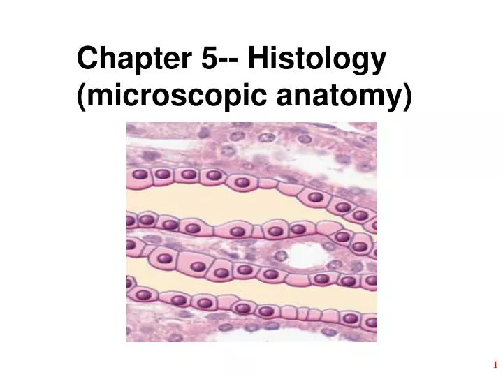
Chapter 5-- Histology (microscopic anatomy). Ch. 5 Study Guide. Read Chapter 5 up to page 170 right before 5.4 Nervous and Muscular Tissue Comprehend Terminology (those in bold in the textbook)

E N D
Ch. 5 Study Guide Read Chapter 5 up to page 170 right before 5.4 Nervous and Muscular Tissue Comprehend Terminology (those in bold in the textbook) Study-- Figure questions, Think About It questions, and Before You Go On (section-ending) questions Do end-of-chapter questions Testing Your Recall— 1-4, 6-10, 13, 17, 18, 20 True or False– 1, 2, 5, 6, 10 Testing Your Comprehension-- #4, #5 2
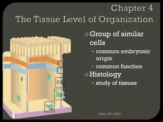
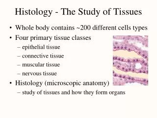
§ The Study of Tissues Whole body contains only 200 different types of cells Four tissue classes (Def. of tissue--?) See Table 5.1. Histology (microscopic anatomy) study of tissues and how they form organs Organ = structure with discrete boundaries composed of 2 or more tissue types Examples:
§ Differences among 4 Tissue Classes Types and functions of cells-- For example: Epithelial, CT, Nervous, Muscular Characteristics of the matrix (extracellular material) Rubbery, stony, or gelatinous Relative amount of space occupied by cells versus matrix CT vs. muscle and epithelium
§ Embryonic Tissues Embryo begins as a single cell divides into many cells that form layers (strata) Three primary germ layers ectoderm (outer) gives rise to: epidermis + nervous system endoderm (inner): mucous membranes: GI tract and respiratory linings; digestive glands. mesoderm (middle) forms mesenchyme (gelatinuous tissue) and then give rise to muscle, bone, and blood
§ Tissue Sectioning (1) 1. Preparation of histological specimens fixation sections mounted on slides & stained 2. Sectioning (slicing) an organ or tissue reduces a 3-dimensional structure to a 2-dimensional slice (see the next 3 slides)
Tissue Sectioning (2) 1 2 3 4 5 • Slices 1 & 5 miss the yolk / cell nucleus • Cell nucleus is smaller in sections 2 & 4 1 5 3 2 4
Tissue Sectioning (3) Image A is a cross section of elbow macaroni, resembling a blood vessel, piece of gut, or other tubular organ. Image B is a longitudinal section of a sweat gland. Notice what a single slice could look like A B
§ Types of Tissue Sections (1) Longitudinal section tissue cut along the longest direction of an organ Cross section tissue cut perpendicular to the length of an organ Oblique section tissue cut at an angle between a cross & longitudinal section
Types of Tissue Sections (2) Would you classify the egg sections as longitudinal, cross, or oblique sections? How would the egg look if sectioned in the other two planes? (Fig. 5.2 question) Practice at home.
§ Epithelial Tissue Introduction (1) One or more layers of closely adhering cells (Top) Forms a flat sheet with the upper (______) surface exposed to the environment or an internal body cavity (Bottom) Sits on basement membrane (basal surface of cells); Fig. X anchors epithelium to underlying connective tissue (Nourishment) No room for blood vessels; . . . CT
§ Epithelial Tissue Introduction (2) Arrangement and Cell Shape— Simple epithelium contains one layer of cells named by shape of cells Stratified epithelium contains more than one layer named by shape of apical cells Fig. 5.3
(Structure) Single row of flat cells (scaly) (Functions) Allows rapid diffusion of substances; secretes serous fluid (Locations) in alveoli, glomerular capsule, endothelium (blood vessels and heart), and serosa (external surface) such as stomach & intestines
§ 2. Simple Cuboidal Epithelium— (Structure) Single row of cube-shaped cells, often with microvilli (Functions) Absorption & secretion; produces mucus (Locations) Liver, thyroid, mammary, salivary and other glands, bronchioles, and most kidney tubules Fig. 5.5
§ 3.Simple Columnar Epithelium— (Structure) Single row of tall, narrow cells vertically oriented, oval nuclei in basal half of cell (Functions) Absorption & secretion; secretion of mucus (Locations) Inner lining of GI tract from stomach to the anus; ducts of gallbladder; uterus, and uterine tubes; some kidney tubes; a few portions of upper respiratory tract
§ 4. Pseudostratified Epithelium— (Structure) Single row of cells not all of which reach the free surface; nuclei at different levels. (Functions) secretes propels mucus (Locations) most of the upper respiratory system from nasal cavity to bronchi; part of male urethra Fig. 5.7
§ Stratified Epithelia Composed of more than one layer of cells & named for shape of __________ cells Deepest cells sit on basement membrane
§ 5A. Keratinized Stratified Squamous Layers of epithelium covered with compact, ______ squamous cells (no nuclei) packed with protein keratin Retards water loss, prevents entrance of organisms Forms epidermal layer of skin (esp. soles and palms) Fig. 5.8 Skin from the sole of the foot
§ 5B.Nonkeratinized Stratified Squamous Multilayered epithelium that lacks surface layer of dead cells forming moist, slippery layer Locations: tongue, oral mucosa, esophagus & vagina Epithelial layer Fig. 5.9 Mucosa of the vagina
Pap smear/test • What? Examination of exfoliated cells • Where? The cervix • Why? • How? Loose cells are examined microscopically for abnormal cells • Who? Between 30-50 years old Fig. 28.5
Fig. 28.5—Pap smears Which one is normal cells?
§ 6. Stratified Cuboidal Epithelium (Structure) Two or more layers of cells; surface cells squareor round (Functions) Secretion and production (Locations) Sweat glands, mammary glands, salivary glands, ovarian follicles, seminiferous tubules Fig. 5.10—Sweat gland ducts
§ 7. Transitional Epithelium (Structure) Multilayered epithelium with rounded (not flattened)surface cells (Functions) Allow stretches and distension (Locations) Urinary tract--part of kidney, ureter, urinary bladder, part of the urethra Fig. X
Qs from Before You Go On (p. 162) Distinguish between simple and stratified epithelia. Explain why pseudostratified columnar epithelium belongs in the former category? Distinguish a stratified squamous epithelium from a transitional epithelium. How do the epithelia of the esophagus and stomach differ? Respective functions?
§ Connective Tissue (CT) Overview Most abundant and variable tissue type 3 structural elements – Consists mostly of (a) G________; (b) F_______ (c) with widely spaced cells Functions of CT: Binding of organs --Ex.a tendon connects muscle to bone Support, protection, movement -- Ex. bones Storage – (energy, electrolytes) Ex. Fats/bones Transport -- Ex. Blood
§ 1. Ground Substance of C.T. Gelatinous or rubbery material found in between cells – Function? Consists of 3 classes of large molecules Glycosaminoglycans (GAGs) – Polysacharides that attract sodium & hold water Ex.-- Proteoglycan is bottlebrush-shaped molecule Forms thick gel that slows the spread of pathogens Cell adhesive glycoproteins Allow themselves bind to matrix elements 36
§ 2. Fibers of C.T. Collagen fibers--called white fibers (Fig. 5.13) Most abundant protein of the body Thick, tough, resist stretch yet flexible Ex. tendons, ligaments & dermis Elastic fibers--called yellow fibers made of E______; recoil like rubberband (elasticity) Ex. skin, lungs & arteries; ability to recoil Reticular fibers Thin collagen fibers coated with glycoprotein Ex. form framework for spleen & lymph nodes 37
Figure 5.13 Collagen Tendons (collagen)
§ 3. Cells of C.T. Fibroblasts -- produce fibers & ground substance WBCs -- wander (mostly in CT) in search of bacteria Macrophages – large phagocytic cells-- arise from monocytes (WBC); function? phagocytosis Plasma cells-- arise from lymphocytes; antibody-producing cells Mast cells – oval shaped; clustered along blood vessels; secrete heparin and histamine Adipocytes or fat cells --store triglycerides
§ Five Types of Fibrous C.T. Divided into 2 broad categories: Loose CT (3 slides followed) contains MORE gel-like ground substance between cells 3 types: A--areolar, B--reticular, C--adipose tissue Dense CT (2 slides followed) FIBERS fill the spaces between cells 2 types varying in fiber orientation: D--dense regular, E--dense irregular 41
§ A-- Areolar Tissue Loose arrangement of collagenous and elastic fibers; scattered cell types; abundant ground substance Locations-- Underlying all epithelia; surrounding nerves, blood vessels, esophagus, trachea Fig. Mesentery
§ B-- Reticular Tissue Loose network of R_________ and cells Forms structural supportive stroma for lymphatic organs Locations-- lymph nodes, spleen, thymus & bone marrow Fig. Spleen
§ C-- Adipose Tissue (Fat) Large, empty-looking cells dominate with thin margins; nucleus pressed against cell membrane; often very pale Functions-- Energy storage, insulation, space filled as cushioning Locations-- Subcutaneous fat beneath skin, breast, heart surface, surrounding organs Fig. 5.18
Figure 5.16b Fig. Adipose tissue
§ D-- Dense Regular CT Structure-- Mainly densely, PACKED, PARALLEL C__________FIBERS; compressed fibroblast nuclei; scanty open space and blood vessels Locations-- Tendons & ligaments Figure 5.16 46
D-- Dense Regular CT Fig. Tendon
§ E-- Dense Irregular CT Densely packed collagen fibers running in ________ directions; scanty open space; few visible cells and blood vessels Function-- Withstands stresses applied in MANY DIFFERENT DIRECTIONS Locations-- Deeper portion of skin; capsules around organs (ex. Liver, kidney etc); sheaths around cartilages and bones Figure 5.17
E-- Dense Irregular CT Fig. Dermis of the skin 49