Download
1 / 33
330 likes | 340 Views
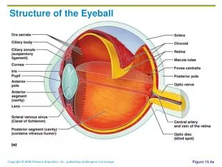
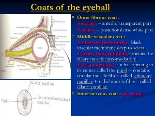
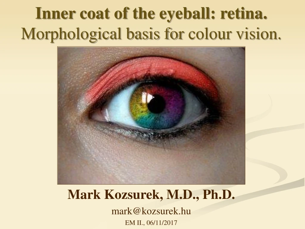
Inner coat of the eyeball : retina. Morphological basis for colour vision. Mark Kozsurek, M.D., Ph.D. mark@kozsurek.hu EM II., 06/11/2017. Inner coat: 1. optic part of the retina 2. nonvisual part of the retina - ciliary part - iridial part. IRIDIAL PART

E N D
Innercoat of theeyeball: retina. Morphological basis for colour vision. Mark Kozsurek, M.D., Ph.D. mark@kozsurek.hu EM II., 06/11/2017
Inner coat:1. optic part of the retina2. nonvisual part of the retina -ciliary part - iridial part
IRIDIAL PART 2 layers of pigmented epithelium CILIARY PART Pigmented epitheliumoutside Non-pigmented epitheliuminside OPTIC PART Pigmented layer (pigment epithelium) Neural layers: 9 layers of neural and glial elements
iridial part ciliary part BLIND PART OF RETINA CORNEA IRIS LENS CILIARY BODY CONJUNCTIVA SCLERA Iridial part of retina consists of two layers of pigment epithelium, while the ciliary part is formed by an outer pigmented and an inner non-pigmented layer.
VITREOUS BODY VISUAL PART OF RETINA L I G H T CHOROID
Cones and rods: the photoreceptors visual pigment: opsin protein + retinal as a chromophor achromatic colour
1. The visual pigments: the opsin family in rods: rhodopsin in cones: short wavelength sensitive opsin in β (blue) or S cones medium wavelength sensitive opsinγ (green) or M cones long wavelength sensitive opsin in ρ (red) or L cones Absorption properties of the visual pigments are exclusively determined by the highly variable opsin peptides, the chromophore is the retinal in all the four types of human photoreceptors!
2. Spectral sensitivity of the photoreceptors Purkinje-effect Rods: much more sensitive, but not selective for the wavelengths (colours). Due to their lower threshold, they work also in the dark. Cones: three types with three different opsins; less sensitive, but selective for wavelength (colour). As they have higher threshold, are active only in light.
daylight, photopic vision (cones) red colours are fading out the cold, blue tones dominate scotopic vision (rods)
rods cones 3. Spatial distribution of cones and rods FOVEA CENTRALIS OPTIC DISK
4. Chemistry and physiology of the fotoreceptors The phototransduction cascade Kramer R H , Molokanova E J Exp Biol 2001;204:2921-2931
Light causes photoisomerization of rhodopsin, activating the heterotrimeric G-protein transducin. The GTP-bound α-subunit activates phosphodiesterase (PDE), which degrades cGMP to GMP. The decrease in cGMP concentration leads to closure of cyclic-nucleotide-gated (CNG) channels, resulting in two effects, a decrease in Ca2+ influx and hyperpolarization of the membrane potential. Lowered intracellular Ca2+ concentration disinhibits guanylate-cyclase-activating protein (GCAP), leading to activation of guanylate cyclase (GC) and resynthesis of cGMP. Photoreceptors are slightly depolarized in dark and hyperpolarize when lit! Illumination decreases or stops the transmitter release!!!
5. Colour vision deficiencies normal rod monochromacy rare no cones at all!
protanopia protanomaly 1% in male 1% in male
deuteranopia deuteranomaly 1% in male 6% in male
tritanopia tritanomaly rare rare
NORMAL DEUTERANOMALY
Why is the default colour of blue? Approximately 10% (!) of male are involved in colour vision deficiences. Most frequently the red and green hues are indistinguishable. Genes encoding the red and green visual pigment proteins are found on the X chromosome, this is why the majority of colour vision deficiencies occur in men. Founder of Facebook, Mark Zuckenberger is green-red colour-blind and as he said, “blue is the richest color for me - I can see all of blue.”
Ishihara Colour Vision Test Plates http://www.youtube.com/watch?feature=player_embedded&v=OkRiz--qexY
„Inverted” Ishihara plate: colours disturb people with normal vision, so they see nothing, but those suffering from colour-blindness can read the text! If the image is converted to grayscale, thus, the disturbing colours are removed, even healthy people can recognize the message!
Visual information processing following phototransduction(phototransduction = conversion of light into neural electric signal)
10. Internal limiting membrane (foot processes of Müller cells) 9. Nerve fibre layer (axons of ganglion cells) 8. Ganglion cell layer 7. Internal plexiform layer 6. Internal nuclear layer (cell bodies of bipolar, horizontal, amacrine and Müller cells) 5. External plexiform layer 4. External nuclear layer (cell bodies of cones and rods) 3. External limiting membrane (foot processes of Müller cells) 2. Neuroepithelial layer (outer parts of rods and cones) 1. Pigment epithelium L I G H T N E U R O N A L S I G N A L Horizontal and amacrine cells are predominantly inhibitory (GABAergic) and contribute to contrast enhancement.
Ganglion cells Ganglion cells Bipolar cells Bipolar cells No convergence Convergence Cones Rods Pigment epithelium Significant convergence (not including the macula and the cones): 5 million cones, 120 million rods, but only 1 million ganglion cells!!!
ON-center and OFF-center cells Receptivefield of bipolar and ganglioncells: a spot withinthewholevisualfieldperceivedbythosephotoreceptorswhichconvergeontheparticularbipolar and ganglioncell. Receptivefieldsconsist of a peripheral and a centralregion.
Blood supply of the retina outer layers: choroid inner layers: central retinal artery - branch of ophthalmic artery of internal carotid artery - superior and inferior, nasal and temporal and macular branches
Must be known for the semifinal: Parts of the inner coat of the eyeball: optic, ciliary and iridial parts of the retina Cell types of the retina: cones/rods, bipolar cells, amacrine and horizontal cells, ganglion cells, Müller cells Layers of the optic retina, regions of interest: the optic disk and the macula/fovea centralis. Basic physiological differences of cones and rods and their spatial distribution. Biochemistry and physiology of vision as well as colour vision defficiences will not be asked!