Download
1 / 54
560 likes | 758 Views
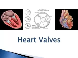
BIOLOGICAL HEART VALVES. Mr. S. V. GHOTKAR. HISTORICAL ASPECT. 1952- The first artificial heart valve to be implanted in humans was a mechanical one implanted by Hufnagel & his attempts heralded the era of cardiac valve prostheses.

E N D
BIOLOGICAL HEART VALVES Mr. S. V. GHOTKAR
HISTORICAL ASPECT 1952- The first artificial heart valve to be implanted in humans was a mechanical one implanted by Hufnagel & his attempts heralded the era of cardiac valve prostheses. 1955- While most workers were working on mechanical devices, it was Murray, who considered replacing a diseased valve with a valve taken from a donor animal. Encouraged by results of his experiments with dogs he implanted a valve harvested from a human cadaver into descending thoracic aorta of a 22 Yr. man who had severe AR. This man was a manual worker & was asymptomatic when seen 6 Yrs later
1956- By this time pump oxygenater became available & Lillehei performed the first aortic valvotomy & repair using retrograde coronary sinus perfusion.These early operations by Hufnagel, Murray & Lillehei had a great significance as they showed that body can tolerate natural & mechanical prostheses as well as native valves were amenable to repair. Here began the competition between these three modalities of treatment.
DEVELOPMENT OF AORTIC HOMOGRAFTS • Subsequently as most researchers were working on mechanical prostheses Murray’s group in Toronto persisted with notion of aortic homograft substitution in orthotopic position.
In 1962 Alfred Gunning & co-workers in Oxford worked out a reliable method for aortic homograft harvest & preparation.
Knowledge of Gunning’s work allowed Ross to perform the first landmark sub-coronary homograft implantation in 1962.This success was soon followed by Barrat-Boyce in New Zealand, Mattias Paneth & Mark O’Brien at Brompton hospital.These were the people who adopted & pioneered homograft aortic replacement at a time when most surgeons preferred the durability & simplicity of Starr Edward valve insertion with a single row of sutures.The homograft insertion was technically more demanding in an era when myocardial preservation was primitive.
To simplify homograft implantation Weldon from John Hopkins hospital published results of laboratory experiments with aortic homograft mounted on a frame.Angell adopted stent mounted homografts for aortic, mitral & tricuspid replacement. Stent mounting increased easiness of implantation & significantly decreased incidence of early post operative regurgitation in comparison to free hand sewn homografts. However, medium term results showed limited durability of stent mounted homograft tissue. The stent thus caused accelerated tissue failure.
DEVELOPMENT OF PERICARDIAL PROSTHESES • Availability of human cadaveric material was always limited & led to search for alternative materials. In Zurich Senning used autologus fascia lata to construct a trileaflet valve, but this turned out to be disappointing.
Marion Ionescu in Leeds mounted heteregounus pericardium on a frame with a sewing ring
DEVELOPMENT OF PORCINE BIOPROSTHESIS • In 1964 after their succes with homografts, Gunning & Duran diverted their attention to preservation of heteregenous valves. In the same year they performed first human stent mounted porcine valve implantation.
In 1965 Duran & Carpentier presented their experience of mercurochrome preserved frame mounted heterografts. This was soon followed by reports by Mark O’Brien about his experience with formalin preserved pig & calf aortic heterografts. However, long term results were poor because of primitive preservation methods.
In 1966 Ionescu developed a stent for mounting aortic heterograts. It had Titanium support, 3 legs covered with dacron velour & dacron felt ring was used as a sewing ring. The porcine aortic valve was sutured within the frame.
Carpentier next produced a stent made of stainless steel coated with Teflon to minimise thromboembolism & he used mercurial salt for tissue preservation. This stent is the precursor of stent used in modern Carpentier- Edward valveWithin 5 yrs of use of stented valves it was realised that stent mounting caused excessive stress on the biological tissue which resulted in accelerated degeneration. This setback caused practice of stent mounting to disappear.
1968 -Preservation of tissues with formaldehyde or mercurochrome was proving disappointing, this problem was soon resolved by Carpentier who employed gluteraldehyde preservation for the first time. The chemical treatment of tissues prevents collagen denaturation & reduces antegenicity of foreign tissues.
Carpentier considered the stent mounted, chemically treated porcine xenograft as a hybrid of biological & mechanical structures & hence named them as Bioprostheses.Central laminar flow was a remarkable positive aspect of these bioprostheses but still presence of the stent was partly obstructive.With efforts of Carpentier, Hancock & Angell gluteraldehyde preserved, stented porcine valves became commercially available & were soon in great demand
DEVELOMPENT OF PULMONARY AUTOGRAFTS In 1967 Ross performed the first pulmonary autograft operation for aortic valve replacement. Initially autogenous pulmonary valve was used as a sub- coronary implant & later as a full aortic root replacement. Ross anticipated that autologus pulmonary valve will remain viable & maintain potential for growth in children. Experience have shown this to be true.
Whilst Ross persisted with his operation others were reluctant to perform a complex double root replacement for isolated aortic disease which otherwise could be treated with very low morbidity & mortality with a stented valve.
DEVELOPMENTS IN REPLACEMENT OF MITRAL VALVE • Ross then attempted to replace mitral valve with a mitral homograft. Though early post-operative hemodynamics were excellent, most patients sustained rupture of chordae tendeniae within a few months of operation. In addition proper placement of papillary muscles inside LV required considerable judgment & skills. Because of these reasons MV homografts replacement has never regained wide spread acceptance.
RESSURECTION OF STENTLESS VALVES • It was being increasingly realised that presence of stent is a disadvantage as it accelerates tissue degeneration. It also tends to obstruct the flow through the valve albeit to much lesser extent than mechanical valves.
In 1988 Tyrone David re-explored the use of stentless gluteraldehyde fixed aortic xenograft. Hemodynamic evaluation showed very small gradients & minimal regurgitation in early implants.
David then went on to develop a low pressure fixed stentless, cloth covered porcine valve. This device is now known as Toronto SPVmarketed by SJM Inc. This device lacks sewing ring & this allows insertion of a larger valve in individual annulus. This valve provided an alternative to aortic homografts which are always in limited supply inspite of development of cryopreservation techniques.
Along the same lines Medtronic Inc. introduced their stentless ‘ Freestyle’ valve in the form of porcine aortic root with Dacron covering of inflow portion. This is a gluteraldehyde preserved, zero pressure fixed valve treated with alpha-amino- oleic acid which acts as a anti-mineralising agent.
SPECTRUM OF BIOLOGICAL VALVES AVAILABLE FOR CLINICAL USE TODAY • Stented porcine valves– e.g. C. E., Hancock • Stentless porcine valves – e.g. Toronto SPV • Pericardial xenografts • Aortic or pulmonary allografts • Pulmonary autografts
STENTED PROSTHESES • Stents used in modern valves are made-up of Elgiloy, Delrin, Acetylco-polymer resin. • Advantages of a stent- Stent provides framework for supporting the leaflets, it also makes implantation easier as compared to valves without a stent.
Disadvantages of the stent • They increase stress on leaflets causing accelerated degeneration. • They are obstructive to flow & hence increase the gradients across the valve. • In mitral position they can cause LVOT obstruction or even rupture of LV especially when a large valve is used in presence of small LV cavity.
Comparison between orifice area in stented and stentless valves Stentless Stented
PRESERVATION/ FIXATION OF TISSUES IN BIOLOGICAL VALVES • Currently gluteraldehyde is the preferred agent. • It cross links the collagen & increases the strength of tissues • It also acts as a sterilizing agent. • Preservation with gluteraldehyde can be done at High pressure ( 60 - 80 mm Hg) Low pressure ( Less than 2 mm Hg ) No pressure ( 0 mm Hg )
Modern prostheses are fixed with either low pressure or zero pressure, this is believed to maintain collagen crimp frame work which will in turn maintain physical properties of biological tissues.
ANTI-MINERALIZATION TRATMENT • Dystrophic calcification is an important factor which contributes to delayed failure of biological valves. This problem is more severe in patients who are young or who have chronic renal failure. • Pathogenesis of bioprosthetic calcification is incompletely understood. • Calcification occurs initially in areas of maximal mechanical stress e.g. commissures. But there are both host related & bioprosthesis related factors.
Gluteraldehyde in itself is a promoter of calcification as it can chelate calcium.There are various agents which are currently being used as decalcification agents v.i.z. Alpha- oleic acid, Polysorbate 80, Sodium decadecyl sulphate, Toludin blue etc.Cells & cellular antigens are also believed to promote calcification. They can be removed by enzymatic detergents leaving only acellular matrix. This process is presently used in SJM-X-CELL investigational prosthesis.
CURRENT RESEARCH IN BIOPROSTHETIC PRESERVATION • Use of gluteraldehyde can be detrimental. There is extensive research going on to find an alternative. • Epoxy compound Denacol has been found to be useful, however high concentrations can be cytotoxic. • Diphosphonates, acyclazide, carbodimide are other agents are under investigation. • Photo-oxidation of bioprostheses promotes collagen cross linking & is being experimented. • Seeding of viable endothelial cell on biological tissues with the aim of endothelization of bioprosthesis is also under investigation.
PROCESSING OF ALLOGRAFTS Presently three types of allografts are available • Cryopreserved • Antibiotic preserved- with cryopreservation they are obsolete now. • Fresh allografts- They are available increasingly due to increased no. of heart transplants.
ALLOGRAFT PROCURMENT • Nondiseased valves are removed from hearts of heart transplant recipients ( Age 6month- 55 years). • Sterile technique is observed during harvesting. • Ascending aorta with aortic valve in situ is removed, origin of innominate artery marks the distal extension & LV base with AML marks the proximal extent of the block.
Harvested sterile graft is then preserved in culture medium ( RPMI 1640) + Antibiotics e.g. Lincomycin + Cefoxitin +Vanco + polymixin B • Stored at 4* C • This graft can then be used directly as a homovital graft or can be cryopreserved (- 40 *C) for future use.
During allograft preservation viability of cellular & mechanical components is very important. • Fresh allografts do have clearly viable cells ( endothelial & fibroblasts ) at the time of implantation. • However, preservation of cellular viability has disadvantage of inducing immunological response
HEMODYNAMICS OF BIOPROSTHETIC VALVE • Bioprostheses have superior hemodynamic performance as compared to any mechanical prosthesis currently available. • Central laminar flow is the the biggest advantage, this minimizes turbulence & also decreases gradients.However, presence of stent does impede flow. • The hemodynamic performance of free hand allograft is excellent & is more or less similar to native valve.
MECHANICAL Vs TISSUE VALVES • OVER THE PAST 20 YEARS MECHANICAL & TISSUE VALVES HAVE CONTINUED TO IMPROVE THOUGH DEBATE OVER THEIR RELATIVE MERITS CONTINUES.
INDICATIONS FOR USE OF BIOLOGICAL VALVES • Biological valves are preferred when - • There is contraindication to use of anticoagulation • Women in reproductive age group who wish to conceive • Patients over 65 year of age
STS database reveals that - Mechanical valves were used in 56-62%, Xenograft in 32-38% & Allograft or autografts in 6%
For AVR, generally 65 yrs & above – Bioprosthesis preferred 40 – 65yrs - Mechanical or allografts 16 – 65yrs – Mechanical, allografts or autografts Paediatric group – Mechanical or autograft
Stentless porcine bioprostheses are currently recommended for elderly with small annuli. It is also proposed as an alternative to allografts. Limited supply of allografts restricts its use to selected patients, particularly those with native valve or prosthetic valve endocarditis complicated by annular abscess or ventriculo- aortic discontinuity. They are also preferred in young & middle aged patients.
CONTRAINDICATIONS TO BIOLOGICAL PROSTHESES • Mechanical valves are preferred than biological in patients who have – - Hypercalcemic syndrome -CRF Bioprostheses are usually avoided in young patients as degeneration & calcification occurs early.
CLINICAL PERFORMANCE OF BIOLOGICAL VALVES • Clinical performance of a valvular prosthesis is judged according to the ‘Guidelines for reporting morbidity after cardiac valvular operations’ ( Edmunds etal, ATS, 1988) Literature provides extensive documentation of individual prostheses, combination of prostheses & comparisons between mechanical & biological prostheses. Only few randomised studies are available which include Veterans Administrative trial (VA) study on valvular heart disease & Edinburgh heart valve trial.
PERFORMANCE OF STENTED XENOGRAFT PROSTHESES Current C-E pericardial prosthesis- Pellerin & co-authors in 64 yr old population report 10 yr freedom of structural failure of 93.5%. At 12 yrs no patient had reoperation for structural failure. Cosgrove & co-authors have come out with similar results.