Download
1 / 1
10 likes | 103 Views
Escherichia coli from healthy animals and food-products of animal origin as reservoir of antibiotic resistance and virulence Souhir Badi 1,2 , Mohamed Salah Abbassi 1 , Paola Cremonesi 3 , Abdennaceur Hassen 2 , Giulia Bignoli 3 , Mario Luini 4 , Bianca Castiglioni 3

E N D
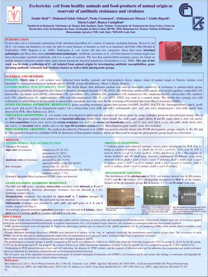
Escherichia coli from healthy animals and food-products of animal origin as reservoir of antibiotic resistance and virulence Souhir Badi1,2, Mohamed Salah Abbassi1, Paola Cremonesi3, Abdennaceur Hassen 2, Giulia Bignoli3, Mario Luini4, Bianca Castiglioni3 1Institut de la Recherche Vétérinaire de Tunisie, Bab Saadoun, Tunis, Tunisia; 2Laboratoire de Traitement des Eaux Usées, Centre de Recherches et des Technologies des Eaux, Technopôle de Borj-Cédria, BP 901, 2050, Hammam-Lif, Tunisia; 3Istituto di Biologia e Biotecnologia Agraria, CNR, Lodi, Italy; 4IZSLER, Lodi, Italy M 1 2 3 4 5 6 fimH 508 bp ibeA 170 bp INTRODUCTION Escherichia coli is a bacterial commensal of the intestinal microflora of a variety of animals, including humans. However, not all E. coli strains are harmless, as some are able to cause diseases in humans as well as in mammals and birds (Dho-Moulin & Fairbrother, 1999; Kaperet et al., 2004). Pathogenic E. coli strains fall into two categories: those that cause intestinalpathologies and those that cause extraintestinalpathologies. Antibiotic resistance in E. coli from human or animal origins has been increasingly reported worldwide, and it is a cause of concern. The fact that antimicrobial resistance genes are found on mobile genetics elements enables their rapid spread among the bacterial population (Guardabassi et al., 2004). The aim of this work was to study a collection of E. coli isolated from animal origins by investigating antibiotic susceptibilities, genes encoding antibiotic resistance and virulence factors as well as their phylogenetic groups. MATERIAL AND METHODS STRAINS: Thirty nine E .coli isolates were collected from healthy animals and food-products (feces, organs, meat) of animal origin in Tunisia. Isolates were identified by classical biochemical methods and by API20E system (BioMérieux, Marcy L'Etoile, France). ANTIMICROBIAL SUSCEPTABILITY TEST: The Kirby-Bauer disk diffusion method was used to determine sensitivity or resistance to antimicrobial agents, according to guidelines developed by the Clinical Laboratory Standard Institute (CLSI, 2010).The followingantimicrobial agents weretested (μg/disc): ampicillin (10), amoxicillin–clavulanicacid (20/10), ceftazidime (30), cefotaxime (30), cefoxitin (30), gentamicin (10), kanamycin (30), streptomycin (10), tetracycline (30), nalidixicacid (30), ciprofloxacin (5), sulphonamides (200) and chloramphenicol (50). E. coli ATCC 25922 wasused as a control strain. The double-disc synergy test withcefotaxime or ceftazidime in the proximity to amoxicillin–clavulanicacidwasused for the screening of Extended-Spectrum Beta-Lactamases (ESBL). GENES ENCODING ANTIBIOTIC RESISTANCE:genesencodingresistanceagainst beta-lactams (blaSHV, blaSHV, blaCTX-M), fluoroquinolones (qnrA, qnrB, qnrS, qepA, and aac(6’)-Ib-cr), kanamycine (aph(3’)-Ia), tetracycline (tetA, tetB, and tetC), sulfonamide (sul1, sul2, and sul3), streptomycin ( strA /strB, aadA) weredetected by PCR. (Ben Sallem et al, 2013) VIRULENCE GENOTYPING:E. coli strains wereinvestigated to determine the presence of variousgenes by using multiplex protocolsdescribedpreviously(Wu et al, 2007). The genestargetedwererelated to (i) bacterialadhesion(fimH, fasA, bfpA, bmaE, iha, nfaE, papC, papGalleles II and III, papGallele I, sfaS, tsh, eaeA); (ii) iron acquisition (fyuA, ireA, iutA); (iii) serumresistance(iss, traT); (iv) toxins and hemolysins (astA, cnf1/2, stx1, stx2, hlyA, cvaA, cdt, cdtB, ehxB, ehxA, eltA, etll, east1); (v) invasion (ibeA, ipaH); and (vi) the type III secretion system (escV). Miscellaneousgene (malX) werealsoincluded. PHYLOGENETIC GROUPING:The method described by Clermont et al. (2000) was used to classify strains into ECOR phylogenetic groups, namely A, B1, B2 and D. This method is based on a multiplex PCR for detection of three genetic markers, which are then used to assign the phylogenetic group based on a flowchart. VIRULENCE GENOTYPING 7 virulence genes were detected amongst various genes investigated by PCR (Fig.3), being (n: number of isolates, %) : fimH (34, 87.1% ), east1(16, 41%), traT (9, 23% ), fyuA (8, 20.5% ), cvaC (2, 5.1%), ipaH (1, 2.5%), and ibeA (1, 2.5% ). The majority of strains harbored only the fimH gene; however, combination of different genes were detected such as: fimH + fyuA + traT + east1 (7 isolates), fimH + ibeA+ traT + east1 (1 isolate), fyuA + traT + east1 (1 isolate), fimH + fyuA + east1 (1 isolate), fyuA + east1 (1 isolate), fimH + traT (1 isolate), fimH + east1 (5 isolate). PHYLOGENETIC GROUPING -The distribution of the phylogroups of 39 E. coli isolates showed that A (21 isolates, 53.8%) and B1 (16 isolates, 41%) phylogroups accounted for 94.8 % of the isolates. Isolates of the phylogenetic groups B2 (2 isolates, 5.1 %) and D (none) were rare. RESULTS ANTIMICROBIAL SUSCEPTABILITY TEST Amongst 39 isolates high rates of resistance kanamycin (34, 87.1%) gentamicin (32, 82%) streptomycin (30, 76.9%) moderate rates of resistance amoxicilline (28, 71.7%) amoxicillin/clav-acide (26, 66.6%) low resistance tetracycline (11, 28.2%) trimethoprim/sulfomethoxazole (10, 25.6%) nalidixic acid (3, 7.7%) Extended-Spectrum Beta-Lactamases (ESBL) were not detected. GENES ENCODING ANTIBIOTIC RESISTANCE The tetA and tetB genes, encoding tetracycline resistance were detected in 8 and 7 isolates, respectively; however, phenotypic resistance was not detected in 3 tet-harboring isolates (Fig.1). Streptomycine resistance was encoded by strpA-strpB in 7 isolates amongst 30 strptomycin-resistant isolates, the aadA gene was not detected. Sulfonamide resistance was encoded by sul1, sul2, and sul3 genes in 1, 4, and 1 isolate, respectively (Fig.2). Interestingly, plasmid mediated quinolone resistance was detected in 6 isolates, where qnrS was in 3 isolates, qnrBin 2 isolates and qnrA in only one. Fig2. Representative gel electrophoresis of PCR sul genes. Line1: 100 bp size marker, Lane 2, sul1 gene, Lanes 3 and 4 isolates with the gene sul2, Lane5, isolate with gene sul3. Fig3. Representative gel electrophoresis of PCR virulence genes. LineM: 100 bp size marker, Lane 2, isolate harboring fimH and ibeA , Lanes 2 to 6 isolate harboring fimH Fig1. Representative gel electrophoresis of PCR gene tetB • DISCUSSION • The finding of high rates of resistance against aminoglycosides and low resistance to tetracycline and trimethoprim/sulfomethoxazoleisuncommon. Indeed, high rates of resistanceagainsttetracyclin and trimethoprim/sulfomethoxazoleisatypicalcharacteristic of E. coli of animal origin, however, lowresistance to aminoglycosides has been mainlyreported • ESBL producer-isolateswere not detected. In Tunisiathisphenotyperemains rare in contrast to the global emergence of E. coli producingESBLsfrom animal whichconstitute a real threat for humanhealth. • PlasmidMediatedQuinolone Resistance (PMQR) weredetected in 6 isolates. In our case, as reportedworldwide, the predomiantegenesremain of qnr-types. The occurrence of such PMQR can not bedetected by antibiogramm but might select for mutations in gyrA and parCgenesleading to highlevel or quinolone resistance • Most strains were considered as commensal because they belong to groups A and B1. • The predominance of genetic groups A and B1 compared to B2 and D was reported by Salhi et al. (2010) who found the following frequencies: 55% for group A, 18.3% for the B1 group, 17.4% for the B2 group and 9.2% for group D. By contrast, Ewers et al. (2007) showed the dominance of group A (46.1%) and B2 (35.1%) compared to group B1 (1.8%) and D (17%). • Virulotyping by PCR showed absence of stx1 and stx2 indicating the absence of STEC isolates, and the majority of detected genes are for adhesion, iron acquisition and serum resistance which are important for Intestinal colonizationand survival during bacterial infection. • The ibeA gene, one of the important invasion-associated genes in neonatal meningitis Escherichia coli (NMEC), was found in one E. coli isolate, this finding is worrisome and highlight the possible dissemination of such very virulent isolate to human. References Ben Sallem et al, (2013). Vector Borne Zoonotic Dis 13:98-102.Clermont et al, (2000). Appl Env Microbiol, 66: 4555-4558; CLSI document M100-S20. Wayne Pennsylvania 2010; Ewers et al, (2007). Int J Med Microbiol. 297(3):163-76; Salehi et al, (2010). Trop Anim Health Prod 42: 1497-1504; Wu et al, (2007). Appl. Environ. Microbiol 73: 83- 91.