Download
1 / 21
210 likes | 325 Views
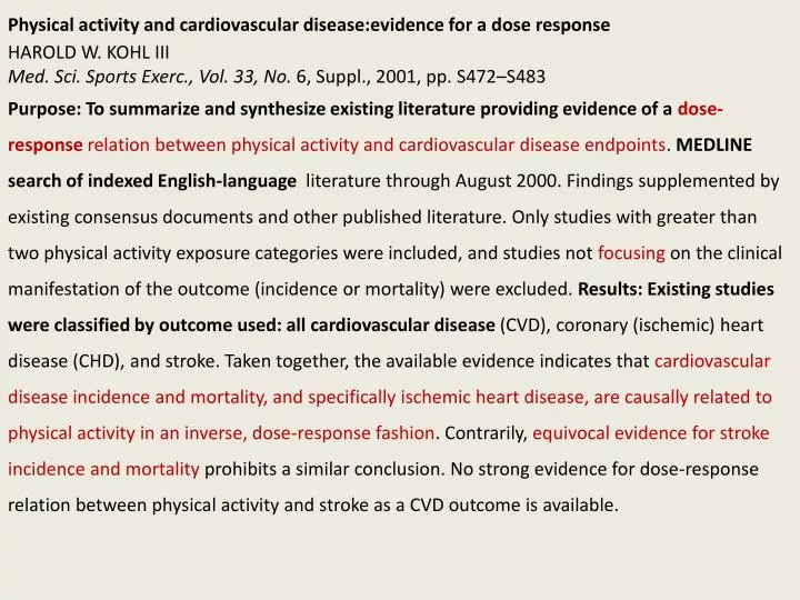
Physical activity and cardiovascular disease: evidence for a dose response HAROLD W. KOHL III Med. Sci. Sports Exerc ., Vol. 33, No. 6, Suppl., 2001, pp. S472–S483

E N D
Physical activity and cardiovascular disease:evidencefor a dose response HAROLD W. KOHL III Med. Sci. Sports Exerc., Vol. 33, No. 6, Suppl., 2001, pp. S472–S483 Purpose: To summarize and synthesize existing literature providing evidence of a dose-response relation between physical activity and cardiovascular disease endpoints. MEDLINE search of indexed English-language literature through August 2000. Findings supplemented by existing consensus documents and other published literature. Only studies with greater than two physical activity exposure categories were included, and studies not focusing on the clinical manifestation of the outcome (incidence or mortality) were excluded. Results: Existing studies were classified by outcome used: all cardiovascular disease (CVD), coronary (ischemic) heart disease (CHD), and stroke. Taken together, the available evidence indicates that cardiovascular disease incidence and mortality, and specifically ischemic heart disease, are causally related to physical activity in an inverse, dose-response fashion. Contrarily, equivocal evidence for stroke incidence and mortality prohibits a similar conclusion. No strong evidence for dose-response relation between physical activity and stroke as a CVD outcome is available.
(The FASEB Journal. 2000;14:1685-1696.) Nitric oxide-mediated metabolic regulation during exercise: effects of training in health and cardiovascular disease BRONWYN A. KINGWELL There is increasing evidence that nitric oxide (NO) is an important hemodynamic and metabolic regulator during performance of physical activity. Furthermore, there are adaptations in this system as a result of exercise training that are likely to contribute to increased functional capacity and the cardioprotective effects associated with higher fitness levels. The production of NO from L-arginine is catalyzed by the dioxygenase, nitric oxide synthase (NOS), which closely resembles cytochrome P450. Three isoforms of NOS, termed nNOS (neuronal), iNOS (inducible), and eNOS (endothelial), have been recognized. NO has been implicated in such diverse processes as vasodilation, inhibition of platelet aggregation, immune function, cell growth, neurotransmission, metabolic regulation, and excitation contraction coupling.
NO AS A METABOLIC REGULATOR DURING EXERCISE Matching tissue oxygen and substrate supply to demand during physical activity is controlled both by blood delivery and the capacity of cells to extract these substrates. As discussed below, NO appears to play a role in both of these processes. Empirical evidence to support a role for NO during exercise includes elevation in exhaled NO as well as increased urinary excretion of the NO second messenger cyclic GMP and the NO metabolite nitrate during performance of exercise in athletes. The mechanisms controlling skeletal muscle blood flow during exercise are complex and involve neural, metabolic, endothelial, myogenic, and muscle pump control. These mechanisms modulate blood flow via effects on perfusion pressure and the caliber of resistance vessels. Traditionally, vessel caliber has been thought to represent a balance between vasodilation mediated directly by production of metabolites from the exercising muscle and sympathetic activation via muscle metabo- and mechanoreceptor stimulation. NO derived from both the endothelium (endothelial NOS, type III) and skeletal muscle (neuronal NOS, type I) may, however, play an important role in matching tissue perfusion to demand.
Stimuli such as adenosine, acidity, temperature, pO2, pCO2, magnesium, and potassium ions contribute to dilation of the microvessels. Other mechanisms mediate upstream dilation of larger ‘feed’ arteries. Vascular shear stress that is determined by blood flow and viscosity is now a well established stimulus for elevation of intra endothelial Ca2+ levels and release of NO from the vascular endothelium. NO formed from this reaction then diffuses to underlying vascular smooth muscle cells, where it activates guanylate cyclase to produce cGMP from GTP and ultimately vasodilation. Thus, microvessel dilation in response to accumulation of vasodilatory metabolites creates a pressure gradient that stimulates flow-mediated dilation of upstream arteries by shear stress-induced release of NO from the endothelium, which permits increased microvascular flow without reduction in muscle perfusion pressure. Skeletal muscle metabolism Both neuronal and endothelial NOS isoforms are constitutively expressed in rat skeletal muscle fibers whereas in humans, nNOS is found in skeletal muscle fibers and eNOS is present in the endothelium of vessels perfusing muscle
NO production from skeletal muscle has been implicated in metabolic control via effects on blood delivery, glucose uptake, oxidative phosphorylation, contractility, and excitation-contraction coupling. NO spares metabolic reserves by promoting glucose uptake and by inhibiting glycolysis, mitochondrial respiration, and phosphocreatine breakdown. The NOS inhibitor L-NMMA infused into the femoral artery during cycling reduced glucose uptake by 48% compared with a control, saline infusion . Oxygen consumption It is well known that large local concentrations of NO produced in response to inducible NOS activation inhibit cellular respiration in a pathophysiological setting. Studies in conscious dogs, however, support the notion that tissue oxygen consumption is modulated physiologically in vivo by constitutively produced NO. The expected reduction in contractility as a result of inhibition of these processes by NO has been observed in the heart and skeletal muscle. These opposing actions of NO on contractile function must be interpreted in the light of studies showing that contraction induces a decline in muscle NOS activity, which if localized to the mitochondria might represent a compensatory mechanism through which muscle contractility and mitochondrial function are protected from the inhibitory influence of NO .
Cardiac muscle function In addition to the role of endothelially derived NO in the coronary vasculature, human cardiac muscle expresses both eNOS and nNOS whereas iNOS is inducible in disease states, including cardiomyopathy. NO appears to inhibit contractile function and oxygen consumption. NO appears to inhibit glucose uptake in the myocardium at rest. The contractile effects are consistent with myocardial relaxation and reduced diastolic tone and are mediated in part by inhibition of respiratory chain enzymes and creatine kinase. Summary NO potentially affects metabolic control during exercise via multiple mechanisms, including: Elevation in skeletal muscle and cardiac blood flow and increased delivery of oxygen, substrates, and regulatory hormones (e.g., insulin); Preservation of intracellular skeletal muscle energy stores by promoting glucose uptake, inhibiting glycolysis, mitochondrial respiration, and phosphocreatine breakdown; Depression of contractile function. Together, these actions of NO on blood flow, substrate utilization, and contractile function appear to be directed toward protection from ischemia.
EFFECTS OF EXERCISE TRAINING ON NO FUNCTION IN HEALTHY INDIVIDUALS From the preceding section it can be seen that NO has multiple roles in the circulatory and metabolic response to an acute bout of exercise. It is not surprising, therefore, that this system adapts in response to training and that such adaptations may contribute to enhanced exercise capacity and reduced cardiovascular disease risk. To date, most studies of the effects of exercise training on NO function have focused on the regulation of vascular tone and blood flow rather than metabolic or other effects. In dog models, exercise training enhanced reactivity to NO-dependent agonists in both proximal coronary arteries and coronary microvessels, but the opposite was true in rats and pigs. There are clear species and regional differences in the NO response to training, highlighting the importance of human studies.
Evidence in humans for chronic changes in the NO system with training is accumulating. Recent work suggests that endothelium-dependent dilation may be altered by training in the rest period between exercise bouts and that the effect may not be restricted to the trained muscle bed. Data indicate first that whole-body dynamic exercise may represent a powerful stimulus for adaptations in the NO system, and second that increased vascular shear stress as a result of elevation in heart rate, pulse pressure, blood viscosity, and blood flow may alter NO function in non exercising muscle beds. Whereas basal NO production appears unaffected at rest by long-term training, acetylcholine-stimulated release is increased, possibly relating to lower total cholesterol in athletes. This effect implies a greater endothelium-dependent vasodilator reserve in athletes, which would increase capacity to perform localized exercise not limited by cardiac considerations.
Summary The vast animal literature together with more recent human studies indicates that endurance exercise training for a period ranging from days to several weeks enhances basal release of nitric oxide from the aorta, active and inactive muscle, and coronary arteries. This adaptation may contribute to the reduction in resting blood pressure that can be observed after as little as 4 wk of training. Increased vascular NO production appears to be a transitory response to training that progresses to structural and other sustained adaptations. Training also enhances agonist-induced, endothelium-dependent dilation in these same vascular beds, but is associated with training durations ranging from weeks to months. Such adaptations would be expected to enhance blood and substrate delivery to cardiac and active skeletal muscle, thus contributing to enhanced exercise capacity.
IMPLICATIONS OF IMPAIRED NO FUNCTION FOR EXERCISE CAPACITY Endothelium Impaired release and/or bioavailability of endothelial NO are associated with a growing list of cardiovascular disease risk factors including hypercholesterolemia, hypertension, smoking, and diabetes and in established coronary disease and cardiac failure. Impaired release may be due to down-regulation of NOS expression or defects in the shear stress or agonist-linked receptor mechanisms that activate NOS. In patients with hypercholesterolemia and coronary atherosclerosis, coronary and systemic arteries constrict during exercise, probably reflecting loss of dilator regulation by the coronary endothelium as a consequence of diminished NO release or increased degradation.
Summary Definitive evidence for a pivotal role of NO in the impaired response to exercise in cardiovascular conditions is not yet available. Furthermore, it is difficult to separate the specific limitations of NO dysfunction on exercise capacity from limitations related to other aspects of disease; however, the data cited and mechanistic plausibility support the contention that NO dysfunction limits exercise capacity. The major mechanism appears to be reduced blood delivery to active muscle including the pulmonary and coronary circulations. These limitations may be particularly important in cardiac failure.
EFFECTS OF EXERCISE TRAINING ON NO FUNCTION IN CARDIOVASCULAR DISEASE The therapeutic potential of training to normalize NO-dependent vasodilation in disease states has been examined in a number of studies, although there are few published studies on NO-related metabolic effects including glucose uptake. With regard to endothelial function, training has the potential to provide benefit via a number of different mechanisms, including: increased shear stress-induced release of NO and prostaglandins; increased expression of endothelial NOS; reduced inactivation of NO by superoxide or other oxygen-derived free radicals. The role of NO in modulating vascular tone after training must be defined in terms of type of training, vascular region, and time course of the training response. NO system is modified by training in the setting of cardiovascular disease and these effects may contribute to increased functional capacity. However, the role of NO in the coronary circulation and skeletal muscle particularly with regard to glucose uptake is yet to be established.
Vagal modulation of heart rate during exercise: effects of age and physical fitness MIKKO P. TULPPO,1,2 TIMO H. MA¨ KIKALLIO,1,2 TAPIO SEPPA¨ NEN,1 RAIJA T. LAUKKANEN,2 AND HEIKKI V. HUIKURI1 1Department of Medicine, Division of Cardiology, University of Oulu, 90220 Oulu; and 2Merikoski Rehabilitation and Research Center, 90100 Oulu, Finland Am J Physiol Heart Circ Physiol 274:H424-H429, 1998.
Fig. 2. HR (A), 2-D vector analysis of Poincare´ plots as indicated by SD1 normalized for average R-R interval (SD1n; B), and high frequency (HF) power of spectral analysis (HF power) normalized for average R-R interval (CCV%; C) in 3 age groups (fitness-matched) during exercise. Values are means 6 SD. Kruskal-Wallis H-tests were used at each exercise intensity level (among all 3 groups) followed by post hoc analysis (Mann-Whitney U-test) between young group and old group. Xx P < 0.01 and xxx P < 0.001 for young group compared with old group. ns, Not significant.
Fig. 4. HR (A), 2-D vector analysis of Poincare´ plots (SD1n; B), and HF power (C) in 3 fitness groups (age-matched) during exercise. Values are means 6 SD. Kruskal-Wallis H-tests were used at each exercise intensity level (among all 3 groups) followed by post hoc analysis (Mann-Whitney U-test) between good fitness group and poor fitness group. X P < 0.05, xx P < 0.01, and xxx P < 0.001 for good fitness group compared with poor fitness group. ns, Not significant.
Physical fitness is related to vagal modulation of HR during exercise independent of aging. This provides further evidence that good aerobic fitness has beneficial effects on cardiovascular autonomic function. Experimental data have shown that vagal activity prevents ventricular fibrillation during exercise and that exercise training confers anticipatory protection from sudden death by enhancing cardiovascular autonomic Function.
Response of blood lipids to exercise training alone or combined with dietary intervention ARTHUR S. LEON, and OTTO A. SANCHEZ Med. Sci. Sports Exerc., Vol. 33, No. 6, Suppl., pp. S502–S515, 2001 Advances in the understanding of the role of blood lipids in atherosclerosis, cause of coronary heart disease (CHD), and related cardiovascular diseases. Specific questions that are addressed in this report : 1) Does the available evidence support the hypothesis that endurance exercise training has a favorable influence on the blood lipid profile relative to future risk of CHD? 2) Does the blood lipid responses to training differ by the study subjects’ sex, age, or race/ethnicity, and baseline lipid levels, and baseline relative body weight and its change with training? 3) Are the lipid responses to exercise related to the intensity, duration, the weekly volume of energy expenditure, the length of the endurance exercise program, and the associated changes with training in maximal oxygen uptake (V˙ O2max)?
COLESTEROLO E LIPIDI: TC: colesterolo totale LDL-C: frazione a bassa densità, considerato il “colesterolo cattivo” HDL-C: frazione ad alta densità, considerato il “colesterolo buono” TG: trigliceridi; la forma di deposito dei grassi animali
The most frequently observed change is an increase in HDL-C, a protective factor against CHD (Evidence Category B). It is estimated that for every 0.026 mmol·L-1 (1 mg·dL-1) increase in HDL-C, the risk for a CHD event is reduced by 2% in men and at least 3% in women. Reduction in TC, LDL-C, and TG also may occur with training. In general, a 1% reduction in LDL-C is associated with a 2–3% lower risk of CHD. Exercise training also appears to attenuate the reduction in HDL-C accompanying a decreased dietary intake of saturated fat and cholesterol to promote reduction of LDL-C. Sex is not a predictor of responsiveness of HDL-C to training, with adult men and women appearing to respond similarly. Age also does not appear to be a predictor of lipid responsiveness to exercise training, with elderly men and women as likely, or perhaps even more likely, than younger individuals to increase HDL-C with training. There have been only a limited number of studies on the effects of different exercise intensities on blood lipids. Most of the studies used an exercise prescription involving moderate- to hard-intensity activities for at least 30 min, three times per week. There also is limited evidence that lower intensity (light-intensity) exercise may be as effective as moderate-intensity exercise in raising HDL-C.