Download
1 / 46
460 likes | 804 Views
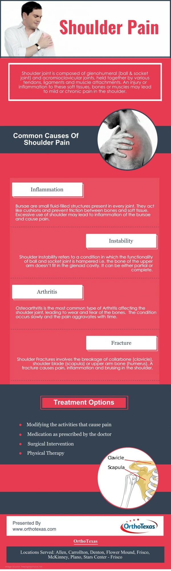
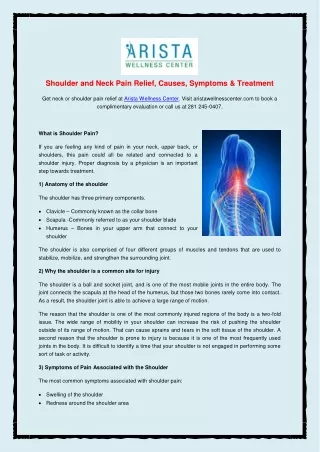
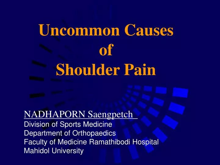
Uncommon Causes of Shoulder Pain. NADHAPORN Saengpetch Division of Sports Medicine Department of Orthopaedics Faculty of Medicine Ramathibodi Hospital Mahidol University. Shoulder pain. Related history Characteristic Location Onset Provocative symptom(s)

E N D
Uncommon Causes of Shoulder Pain NADHAPORN Saengpetch Division of Sports Medicine Department of Orthopaedics Faculty of Medicine Ramathibodi Hospital Mahidol University
Shoulder pain • Related history • Characteristic • Location • Onset • Provocative symptom(s) • Other possible correlated symptom(s)
Differential diagnosis • Lethalgy medical causes : MI, PE • Neurological : poliomyelitis, C-spine diseases (disc, root, cord) • Neuropathy : neuralgic amyotrophy, mononeuritis multiplex • Entrapment : brachial plexus, peripheral nerves • Thoracic outlet syndrome • Primary shoulder disorders*
Neuralgic amyotrophy • Brachial plexus neuropathy • Brachial plexitis • Parsonage-Turner syndrome • (136 pts Lancet 1948;1:973-8) • Idiopathic brachial neuritis • Serum neuritis • Shoulder girdle syndrome • …… • …… • Neuralgic amyotrophy
Neuralgic amyotrophy • Incidence 2-3 : 100,000 • (McDonald BK Brain 2000;123:665-76) • (Beghi E Ann Neurol 1985;18:320-3) • The ever largest series 246 pts • (van Alfen N Brain 2006;129:438-50) • Male > female (2:1) • Etiology : • immune-mediated process • infection (25-55%) • strenuous exercise, vaccination, • post-surgical, hereditary • (Sathasivam S JBJS Br 2008;90:550-3)
Clinical features • Pain is the first symptom (90%) • Acute severe burning shoulder pain, radiating to the arm ~39.7% • (van Alfen N Brain 2006;129:438-50) • Worsen by movement : most com fort in adduct shoulder and • flex elbow • Not aggravated by Valsalva maneuver and Spurling’s test • (DDx from C-radiculopathy)
Important feature • Pain subsides and follows by muscle weakness** • Mimic rotator cuff/deltoid tendinopathy….that needs to be proved with imaging studies!
Clinical features • Weakness : develops within 2 wks, affects the upper brachial plexus 50% (isolation or several involvement) • (Hawkins RH JBJS Br 1987;69:195-8) • Inferior subluxation of the HH • Sensory involvement : hypoesthesia at deltoid, lateral arm and radial forearm • Autonomic dysfunction : trophic skin, increase sweating, etc.
electromyography • Best done after 3 wks of the onset • Fibrillation potential • Abnormal distal conduction velocity • (Flaggman PD Arch Neurol 1980;37:160-4) • Demyelination in early stage • (O’Brien MD Lancet 1980;ii:975) • Axonal degeneration • Delayed distal latencies and decrease amplitude of CMAP • (Mullins GM Neurol Neurosurg 2007;109:661-6)
Other investigations • CXR : useful to R/O Pancoast tumor • Blood tests : LFT, ESR, CRP, RF, ANA and anti-dsDNA • CSF study : mild pleocytosis, oligoclonal bands
Imaging studies • Coronal T1-weighted conventional spin echo (TR 700 ms, TE 20 ms) • Slide thickness 3 mm • MRN is superior to conventional MRI during the acute stage of diagnosis of NA • (Duman I Neurologist 2007;13:219-21)
MRI / MRN MRI Conventional post-Gd, Coronal view MRN Coronal short tau inversionrecovery view
treatment • Steroid : methylprednisolone • Narcotic : hydrocodone • NSAIDs • Muscle relaxant : cyclobenzaprine • (Miller JD Am Fam Physician 2000;62:2067-72) • Physiotherapy & exercise
Prognosis • Good prognosis with recovery onset 1-3 yrs. • (McCarthy EC CORR 1999;368:37-43) • 75 % recover within 2 yrs • (Tsairis P Arch Neurol • 1972;27:109-17)
Suprascapular nerve Entrapment (SSNE) • Usually been missed until atrophy or fatty infiltration are seen • Even miss by plain x-ray or CT scan • EMG is the gold standard to diagnose • Imaging hints : muscular edema, atrophy and fatty change • Labral/spinoglenoid notch cyst
Location of ssne Superior transverse Scapular ligament Inferior transverse Scapular ligament (Spinoglenoid lig.)* (Westerheide KJ Orthop Clin North Am 2003;34:522)
Suprascapular nerve entrapment(SSNE) • Suprascapular notch –SSN (SST+IST) • traction /compression • tethering effect of the ligament • Spinoglenoid notch –SGN (IST alone) • traction (overhead athlete) • a ganglion cyst*
Muscular edema is the most sensitive for SSNE compares with EMG (Ludig T Eur Radiol 2001;11:2161-9)
Symptomatic ganglion cyst • Non operative • avoid repetitive overhead • PT scapular stabilizers and cuff • Operative • image guided aspiration (?recur) • open decompression • arthroscopic decompression • or combine procedures
Amber-color, gelatinous material • Postero-superior • capsulotomy • Repair of type II • SLAP lesion • (Abboud JA CORR 2006) • (Iannotti JP Arthroscopy 1996) • (Lichtenberg S Knee Surg Sports Traumatol Arthrosc 2004)
42 pts with S-P cyst and labral tear • Capsulolabral tear forces joint fluid into the tissue => one-way valve cyst • Labral repair alone leads to cyst resolution and pain relief • Average cyst diameter 2.4 cm • No attempt to evacuate the cyst • Improved Rowe score and MRI
Pre operative Post operative Persist TMi atrophy ( Schroder CP JBJS Am 2008;90:523-30)
Working room under the SST Portal localization (J Shoulder Elbow Surg 2008;17:616-23)
Arthroscopic decompression 2 notches at the same time (fresh cadavers)
Satisfied decompression Complete decompression 18/20 of suprascapular notch 20/20 of spinoglenoid notch
Facts • First reported in 1955 and1983 • (Cahill BR J Hand Surg Am 1983;8:65-9) • Fibrous band compress the axillary nerve and posterior circumflex humeral artery • Vague posterior shoulder pain, paresthesia and weakness of TMi &posterior deltoid • 0.8% incidental finding from MRI • (Cothran RL Jr Am J Roentgenol 2005;184:989-92)
The fibrous sling arises from the LHT is a normal finding (McClelland D J shoulder ElbowSurg 2008;17:162-4)
Provocation tests • Shoulder abduction to 90º • fully internal rotation…then • external rotation* • Subsequent pain and paresthesia over the shoulder blade (McClelland D J Shoulder ElbowSurg 2008;17:162-4)
Investigations for QSS • MRI provide additional detail to electrophysiologic studies • acuteT2 fast spin echo FS, • increase SI = neurogenic edema • chronicT2 spin echo, diffusely increaseSI = outline muscle bulk • (Breddella MA Skeletal Radiol 1999;28:567-72)
Doubt MRI (Sofka CM Skeletal Radiol 2004;33:514-8) • 2,563 shoulder MRI with 3% isolated TMi denervation (some with EMG) • Other causes of TMi atrophy • surgical intervention posterior portal malposition, instability sx, capsular thermoplication • translation/dislocation traction, capsulolabral damage • nerve irritation RC, LHT
5 years later (Sanders TG Arthroscopy 1999:6;632-7)
Other Anatomic causes • Glenoid labral cysts • A ganglion • Muscle (SSC) hypertrophy • A spike of bone after a scapular fracture
Surgical decompression (McAdams TR Am J Sports Med 2008;36(3):528-32) 3-D CT angiogram