Download
1 / 19
200 likes | 226 Views
Chapter 46 Respiratory Monitoring and Management of the Patient in the Intensive Care Unit. Objectives. Discuss the principles of monitoring the respiratory system Discuss the risks and benefits of intensive care unit (ICU) monitoring techniques.

E N D
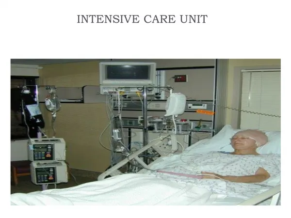
Chapter 46Respiratory Monitoring and Management of the Patient in the Intensive Care Unit
Objectives • Discuss the principles of monitoring the respiratory system • Discuss the risks and benefits of intensive care unit (ICU) monitoring techniques. • Discuss why the caregiver is the most important monitor in the ICU. • Describehow to evaluate measures of patient oxygenation in the ICU.
Objectives (cont.) • Define why Paco2 is the single best index of ventilation for critically ill patients. • Describe the approach used to evaluate changes in respiratory rate, tidal volume, minute ventilation, Paco2, and end-tidal Pco2 values for monitoring purposes. • Discuss monitoring techniques used in the ICU to evaluate lung and chest wall mechanics and work of breathing. • Discuss the importance of monitoring peak and plateau pressures in patients receiving mechanical ventilatory support.
Objectives (cont.) • Describe the approach used to interpret the results of ventilator graphics monitoring.
Introduction to Monitoring • Continuous monitoring or periodic checks • Gray area between diagnostic and monitoring procedures • Risk/benefit ratio
Monitored Values • All data must be evaluated in context of overall clinical presentation. • Instrument inaccuracyrecalibrate • Artifacts • Factitious results: true but temporary (cough) • Treat the pathology, not the errant number. • All values monitored must be considered in relation to what pathology has altered them and how best to treat the pathology.
Monitoring Oxygenation • Tissue oxygenation depends on CaO2 (PaO2 and SaO2), cardiac output, and oxygen uptake • Pulse oximetry (“fifth vital sign”) • Provides noninvasive measurement of SaO2, referred to as SpO2 • Monitors only oxygen, not ventilation • Significant limitations
Other Oxygen Indices • Oxygen consumption • Difficult to measure, so seldom used • Normal 250 ml/min, 25% of oxygen delivery • P(A a)O2 • Healthy patient • 21% O2, gradient is 5 to 15 mm Hg • 100% O2, gradient is 100 to 150 mm Hg • An abnormal increase is associated with gas exchange problems.
Other Oxygen Indices (cont.) • PaO2/FIO2 ratio (P/F ratio) • Normal P/F ratio is 400 to 500. • In ALI, this falls below 300. • In ARDS, it will be < 200. • Most reliable index of gas exchange if FIO2 > 0.50 and PaO2 < 100 mm Hg • QS/QT (physiologic shunt) • Increased if pulmonary venous admixture occurs (mixed venous blood exits A/C membrane unchanged)
Monitoring Ventilation • Routine monitoring includes • PaCO2, which defines adequacy of ventilation • T, f, and E • Low VT and high f often indicate distress • VD/VT • Normal 0.20 to 0.40 • Higher ratio indicates more wasted ventilation • ICU common to be >0.70 • >0.60, patient is unlikely to sustain spontaneous ventilation . . V V . .
Compliance • Compliance is ΔV/ΔP or effective VT/(Pplat PEEP) . • Normally, this is 60 to 100 ml/cm H2O . • In severe ARDS, it may be <25 ml/cm H2O . • Many pulmonary diseases alter compliance. • See Box 46-7.
Resistance • Resistance (Raw) = (PIP Pplat)/flow • Normally 1 to 2 cm H2O/L/sec • Intubated, probably 5 to 10 cm H2O/L/sec or more • See Box 46-7 for diseases that alter Raw.
Auto-PEEP • If exhalation is incomplete, auto-PEEP occurs. • Causes ⇑FRC and mean alveolar pressure • Often causes patientventilator asynchrony • Ways to decrease auto-PEEP • Decrease VE • Increase ET • Decrease IT .
Auto-PEEP (cont.) • Adding extrinsic PEEP may overcome the trigger sensitivity issue and facilitate lung emptying. • Slowly increase in 1-2 cm H2O increments till either: • Patient can trigger the ventilator • Auto-PEEP increases
Measuring Auto-PEEP • Presence noted by expiratory flow at the end of expiration • Measured by • End-expiratory hold: most common method • Allows alveolar pressure to equalize with the ventilator pressure. • Esophageal balloon • Increase PEEP until end-expiratory flow is zero • PEEP applied estimates auto-PEEP
Monitoring Breathing Effort and Pattern • P0.1 assesses ventilator drive • Occlusion pressure 100 ms after initiation of inspiration • <6 cm H2O is indicative of patient’s ability to wean from MV. • RSBI (f/VT) • Respiratory muscle fatigue tends toward rapid shallow breathing. • RSBI < 100 indicates patient likely to wean from MV • The lower the RSBI the better
Monitoring Breathing Effort and Pattern (cont.) • Vital capacity (VC) • Effort dependent • VC less than 10 to 15 ml/kg, need for MV • Maximal inspiratory pressure (MIP) • Not effort dependent, as prolonged occlusion of airway stimulates maximal effort • More negative is better • 20 to 30 cm H2O acceptable
Monitoring During Lung Protective Ventilation • Commonly used for ALI/ARDS patients to avoid ventilator-induced lung injury (VILI) • Three principles confirmed • Limit Pplat to <30 cm H2O • Reduce VT to 6–8 ml/kg • Use adequate PEEP to avoid opening/closing injury • Permissive hypercapnia is often used as a lung protective strategy avoid VILI.