Download
1 / 20
200 likes | 379 Views
Case # 2. Clinical progress: 2009 HLA typing: no sibling match Hb S: 27-40% anti-S, anti-Jk(b) TCD velocities conditional range: < 200 cm/s Liver iron content (MRI): 6.7 mg Fe/g RBC transfusion # 16 Commenced Deferasirox (oral Fe chelator) 2010 Hb S: 16-28%

E N D
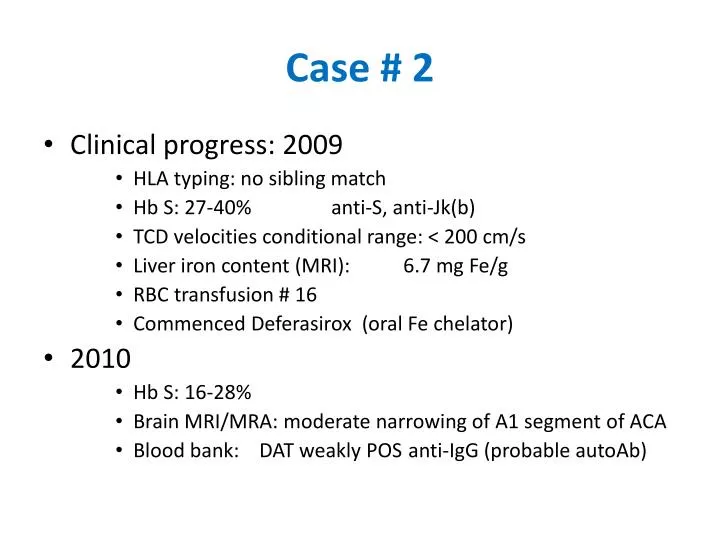
Case # 2 • Clinical progress: 2009 • HLA typing: no sibling match • Hb S: 27-40% anti-S, anti-Jk(b) • TCDvelocities conditional range: < 200 cm/s • Liver iron content (MRI): 6.7 mg Fe/g • RBC transfusion # 16 • Commenced Deferasirox (oral Fe chelator) • 2010 • Hb S: 16-28% • Brain MRI/MRA: moderate narrowing of A1 segment of ACA • Blood bank: DAT weakly POS anti-IgG (probable autoAb)
Transfusion in Patients with Haemoglobinopathies(Sick Kids approach) • Thalassemia (including congenital anemia like Diamond Blackfan Syndrome) and sickle cell disease • Extended red cell phenotyping before first transfusion: • Rh (C, E, c, e), K1(Kell), Fya, Jka, (Jkb, S in SCD patients) • Patient already transfused • Hypotonic saline (0.3% NaCl will lyse normal cells but not sickle cells) AABB Technical Manual 15th edition Method 2.16 • Molecular genotyping • Family studies • Prophylactic antigen matching: • Thalassemia: K negative • SCD: Rh and K matched, Fya, Jk, (S) if alloimmunized (No need for prophylactic Fyb matching • Why not Fyb?
Fy(a-b-) Phenotype • Frequency • Very rare (0%) in Caucasians and Asians • 68% in Africans • Mutation in the promoter region of FYB (–33 T>C), which disrupts a binding site for the erythroid transcription factor GATA-1 and results in the loss of Fy expression on RBCs. • Because the erythroid promoter controls expression only in erythroid cells, expression of Fy proteins on endothelium is normal in people with Fy(a–b–) RBCs. • To date, all blacks with a mutated GATA box have been shown to carry FYB, therefore Fyb is expressed on their nonerythroid tissues. • This explains why Fy(a–b–) individuals make anti-Fya but not anti-Fyb. Immunohematology 2004;20:37-49
The Duffy Antigen/Receptor Chemokine locus • Comprised of 2 exons, spanning ~2 kbp in the region of chromosome 1q22-23 • The single gene is responsible for the expression of Duffy antigens on RBCs and other non-erythroid tissue • Promoter GATA-1 box mutation is responsible for the Fy(a-b-) RBC phenotype (pseudo-null) • The gene product has no obvious function on the surface of RBCs; null RBCs appear to function normally • FY affords protection from malarial invasion • It has been proposed that the gpFy modulates chemokine levels in the blood; regulation of inflammation by scavenging chemokines
Allelic variants of DARC GATA-1 FY*A/FY*B FY*X (wt) FYB ^ ^ ^ -33T 265C 125A FYA ^ ^ ^ -33T 265C 125G (amorph) FY ^ ^ ^ -33C 265C 125A FYX ^ ^ ^ -33T 125A 265T (null) FY0 ^ ^ ^ -33T 265C 125A
Transfusion in Patients with Haemoglobinopathies(Sick Kids approach) • Start with SAGM units (prestorage leukoreduced) • 15 to 20 ml/kg, SAGM red cells have hct approx 0.6 • SCD: Units tested by sickle test (sickle negative for exchange transfusions) • Repeated allergic reactions: pre-med • Recurrent allergic reactions: plasma-reduce • Frequent allergic reactions: Washed red cells from CBS: 24 hours shelf-life • Currently, no Sick Kids patients on washed cells
Case #3 • Female, born in 1980, sickle cell disease • B pos, C-E-c+e+, K-, Fy(a-b-), Jka(a+b+) • 1998: no antibody, transfused 3 units • 1999: anti-K, anti-C, anti-E, autoantibody • Sept 1, 2004, transfused 2 units, O neg, C- E- K- S- Fya-, crossmatch compatible • Sept 14, 2004, 3 units B pos, C- E- K- S- Fya-, crossmatch incompatible
Case #3 (2004) • Anti-IH • 4+ with group O cells by Sal I.S., 370C and IAT • 1-2+with group B cells • Negative to weak with Oh (Bombay) cells • Testing with Oh cells
Anti-IH • I antigen: subterminal portion of the oligosaccarides that are eventually converted to H, A, and B antigens • Most normal adult RBCs are I-positive • H antigen is the substrate for A and B antigens • H antigen expression: O > A2 > B > A1B > A1 > A1B • The most common cold autoagglutinins are directed against the Ii blood group • The most commonly encountered cold autoagglutinin recognizing complex ABH-Ii antigens is anti-IH • Anti-IH does not generally interfere in pretransfusion testing done at 370C, but may be picked up in MTS-gel • Anti-IH is usually not clinically significant, anti-IH causing hemolytic transfusion reaction is very rare but has been reported. Transfusion 2000:40;828
Case #3 (2005-2006) • Anti-IH not detectable • Autoantibody • Recommended for transfusion: group B, C- E- K- M- Fya- • May 5, 2006, transfused 2 units O pos, C- E- K- Fy(a-b-) S-, MTS compatible, Hb 70 to 99 g/L • May 15, 2006, Hb to 50g/L • Strongly reactive with all cells tested, except for 2 Group B Rhnull cells • Episode of hyperhemolysis, eventually recovered
Antibodies to high prevalence antibodies • Red cell alloantibodies • k, Kpb, Lub, Jsb • Jk3, U • African ethnicity: SsU, Jsb, Ata (Augustine), Hy (Holley), Joa (Joseph) • Phenotype patient’s RBCs (Rh and others) • Antigen negative cells • Enzymes and chemicals • Antibodies to reagent/preservative • ABH antibodies • Group O patient, think Bombay or para Bombay • Non group O patient, think IH (more common in A1, A1B, less common A2, B, A2B)
Summary • Phenotype patient before 1st transfusion • If transfused, hypotonic saline for SCD, molecular genotyping and family studies • Autoantibodies are common • SCD patients can make unusual alloantibodies • When investigating for high incidence antibodies, do not forget ABO and reagents • Clinical information including patient’s ethnic background
References • Guidelines for Antibody Investigation. AABB 2010 • Judd’s Methods in Immunohematology, 3rd edition, AABB Press 2008. • The Blood Group Antigen Facts Book, 2nd edition, 2004, Reid & Lomas-Francis, NYBC • Applied Blood Group Serology 4th edition. Issitt 1998.