Download
1 / 23
480 likes | 2.97k Views
Hypoxia: types & effects, Cyanosis, Hypercapnia , Dyspnea , Asphyxia. By Dr. Roomi. TYPES OF HYPOXIA:. There are 4 types of hypoxia: Hypoxic Hypoxia Anemic Hypoxia Stagnant / Ischemic Hypoxia Histotoxic Hypoxia. HYPOXIC HYPOXIA:. Causes :

E N D
Hypoxia: types & effects, Cyanosis,Hypercapnia, Dyspnea, Asphyxia By Dr. Roomi
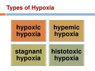
TYPES OF HYPOXIA: There are 4 types of hypoxia: • Hypoxic Hypoxia • Anemic Hypoxia • Stagnant / Ischemic Hypoxia • Histotoxic Hypoxia
HYPOXIC HYPOXIA: Causes: • High altitude decreased PO2 in atmospheric air. • Respiratory muscle paralysis. • Obstructive lung disease (COPD). • Restrictive lung disease (pulmonary fibrosis, pneumothorax). • Depression of respiratory centre (disease/ drug). Effect on arterial PO2: • Decreased arterial partial pressure of oxygen. MCQ • In other types of hypoxia, PO2 is normal.
Clinical features of Hypoxic Hypoxia: • Due to decrease arterial PO2 Interstitial cells in peritubular capillaries of kidney secrete Erythropoietin polycythemia • Hypoxia pulmonary V.C Pulm. Hypertension Rt. Vent. Hypertrophy Rt. Vent. Failure. TREATMENT: O2 treatment is most effective in this type of hypoxia.
ANEMIC HYPOXIA: • Arterial PO2 is normal but inadequate O2 carrying capacity of blood. • Causes: • decrease in Hbconc. • abnormal Hblike met-Hb or Hb-S • CO poisoning: binding site of Hb for oxygen is not available
CO Poisoning • CO is produced by incomplete combustion of carbon. • CO is a colorless & odourless gas. • Hbhas 250 times more affinity to bind with CO as compared to O2. • Carbon monoxide Hb shifts the oxy-Hb curve to left O2 dissociation becomes difficult. • CO also inhibits cytochrome. • In CO poisoning, skin is cherry red colored. • There is no stim. of resp. centre, because arterial PO2 is normal. • When there is 70% carbon monoxyHb in blood death occurs.
Treatment of CO Poisoning: • Remove the subject from source of exposure. • 100% oxygen therapy can help. • Hyper-barric O2can help (O2 with increased pressure = 2-3 atm)
STAGNANT / ISCHEMIC HYPOXIA: CAUSES: • Decreased cardiac output / sluggish blood flowdue to: • heart failure, • hemorrhage, • circulatory shock and • venous obstruction. • EFFECTS: • Blood remains in tissues for longer time, so tissue extracts increased oxygen from blood more AV difference of oxygen concentration. • So, PCO2 increases, it facilitates unloading of oxygen from hemoglobin (shifts the oxy-hemoglobin association dissociation curve to right).
HISTOTOXIC HYPOXIA:(poverty amongst plenty) DEFINITION: Inability of the tissues to utilize oxygeninspite of normal arterial PO2 and oxygen carrying capacity. CAUSES: • Cyanide poisoning (it inhibits cytochromeoxidases oxidative process is inhibited). • Narcotic overdosage(it inactivates the enzyme dehydrogenase inhibition of tissue oxygenation). • Beri-beri (it is deficiency of thiamine co-enzyme which is required for many oxidative reactions). TREATMENT: • Methylene blue or nitrites. These convert hemoglobin met-hemoglobin. • Cyanide + met-hemoglobin cyn-met-hemoglobin (non-toxic compound).
CYANOSIS: • Definition: Bluish discoloration of skin& mucus membrane, when conc. of deoxy-Hb in small blood vessels like capillaries > 5 g/dl.
1. Peripheral: Seen on: exposure to moderate cold & in case of stagnant hypoxia. Seen in: fingers, outer surface of lips. Arterial PO2 remains normal. Types of Cyanosis:
Types of Cyanosis: 2. Central: • Seen in : • case of Congenital heart diseases & • chronic lung disease. • Mostly Arterial PO2 is below normal (due to hypoxic hypoxia).
Conditions in which Cyanosis does not occur: • Severe anemia (less than 5 gram deoxyHb/dl) • CO poisoning (masked due to cherry red complexion) • Met-Hemoglobinemia (chocolate brown discoloration)
DYSPNEA = Air Hunger Dyspnea: shortness of breath (SOB), or air hunger, is the subjective symptom of breathlessness. 3 factors that cause the sensation of dyspnea: 1) Abnormality of respiratory gases in body fluids (mainly hypercapnia & partly hypoxia) 2) Increase work of breathingby respiratory muscles to breath forcefuly e.g. in asthma 3) State of Mind (neurogenic/emotional dyspnea) • More enhanced in people who are claustrophobic (fear of not being able to receive a sufficient quantity of air e.g., small or crowded places).
HYPERCAPNIA: DEFINITION: • Excess CO2 in body fluids. • (Hypercapnia + Hypoxia): Only when hypoxia is caused by hypoventilation or circulatory deficiency.
Causes of hypoxia + hypercapnia (simultaneously): • In hypoxia due to hypoventilation, CO2 transfer between alveoli & atmosphere is affected as much as is oxygen transfer. • In circulatory deficiency decreased blood flow decreased removal of CO2 from the tissues tissue hypercapnia + hypoxia. • But transport capacity of blood for CO2 is more than 3 times that for O2, so resulting tissue hypercapnia in much less than tissue hypoxia.
Severe hypercapnea • When alveolar PCO2 rises above about 60-75 mm Hg air hunger / dyspnea becomes severe. • If PCO2 rises to 80-100 mm Hg lethargy, +/- semicomatose • If PCO2 rises to 120 to 150 mm Hg + / - anesthesia & death • At such high PCO2 Resp. Centre is depressed rather than stimulated vicious circle
ASPHYXIA: DEFINITION: Simultanoeus acute hypoxia & hypercapnia. CAUSES: • Acute airway obstruction • When a person is forced to re-breathe his own air in a confined space.
Mechanism: • During asphyxia hypoxia + hypercapnia strong stimulation of respiratory centre & violent inspiratory efforts heart rate increases, BP increases, CATS increase from adrenal medulla (increase in nor-epinephrine > epinephrine) unconsciousness, convulsions & decrease in respiratory rate death.
O2 THERAPY: (3 ways) Intra-nasal tube O2 mask on nose O2 tent (newborn)
Oxygen therapy is helpful in: • Most helpful in hypoxic hypoxia. • May be helpful in cyanide or CO poisoning • May be helpful in case of Gas Gangrene. • Note: No use in Anemic & Ischemic (stagnant) hypoxia.
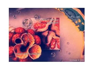
OXYGEN TOXICITY If 100% O2 treatment is given for over 8 hours: If 100% O2 treatment is given for 24-48 hrs: Toxic effects start like: lung damage, decreased ability of alveolar macrophages to kill bacteria, decrease in surfactant secretion, cyst formation in lung may occur, Retrolentalfibroplasia in infants kept in O2 tents for long. Features of airway irritation • sore throat • substernal distress • nasal congestion • coughing
Cause of toxic effects of oxygen: • When O2 is given for longer period toxic effects. • Formation of certain free radicals: • Super-oxide ions (O2-) • Hydrogen peroxide (H2O2) • Prevention of toxic effects of oxygen: • By anti-oxidants like vitamin E.