Download
1 / 1

E N D
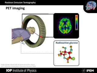
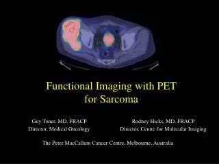
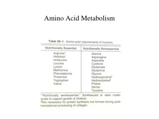
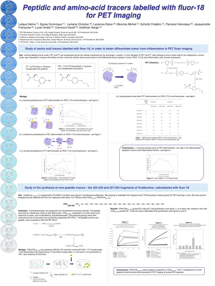
Aim : Among labelled amino-acids, FET and FT are transported across the cellular membrane by the exchanger l–system. In vitro utilisation of FET and FT, after preload or prior to after load of non-radioactive L-amino-acids, was evaluated to measure the effects of intra- and extra cellular amino-acid content on the differential tracers uptake in tumor (ROS 17/2.8) and inflammatory cells (human leukocytes). Study of amino acid tracers labelled with fluor-18, in order to better differentiate tumor from inflammation in PET Scan imaging FET Chemistry : FET : O-(2-[18F] fluoroethyl)-L-Tyrosine : not incorporated into proteins FT : 2-[18F] fluoro-L-Tyrosine : incorporated into proteins d) L-phenylalanine load after FET administration on ROS 17/2.8 and leukocytes : see table 1 Biology a) L-tyrosine preload prior to FET administration on ROS 17/2.8 and leukocytes : see figure 1 Figure 1 : preload effect = ratio between the tracer uptake into preloaded cells and control cells b) L-methionine preload prior to FET administration on ROS 17/2.8 and leukocytes : see figure 1 Table 1 : differenciation between ROS 17/2.8 and leukocytes = ratio between FET content in ROS 17/2.8 and in the inflammatory cells Conclusions :L–tyrosine preload, prior to FET administration, can help in the differentiation between tumours and inflammatory lesions : see figure 3 c) L-tyrosine preload prior to FT administration on ROS 17/2.8 and leukocytes : see figure 2 Study of the synthesis of new peptidic tracers : the 323-339 and 257-264 fragments of Ovalbumine, radiolabeled with fluor-18 Figure 2 : preload effect = ratio between the tracer uptake into preloaded cells and control cells Figure 3 : tracer uptake = % of loaded activity Aim : Ovalbumin 323-339 is recognised by DC MHC II complex and induces T-lymphocyte proliferation. We propose to radiolabel this fragment with [18F]Fluoroethyl to allow pulsed-DC PET-tracking in mice. We here present biological results obtained with the non-radioactive derivative, N-[19F]fluoro-ethyl-OVA323-339 (FEthOVA323-339). OVA 323-339 : NH2– Ile – Ser – Gln – Ala – Val – His – Ala – Ala – His – Ala – Glu – Ile – Asn – Glu – Ala – Gly – Arg – OH Chemistry : fluoroethyltosylate was prepared from fluoroethanol and tosyl chloride. The peptide was build on solid phase made up with Wang resin. OVA323-339, protected on its side chains and carboxylic function, was N-alkylated by fluoroethyltosylate. The protecting groups were then removed and the final product was purified by preparative RP-HPLC. The peptide and the neo peptide, were anaylsed on MS and RP-HPLC. Figure 1 : Figure 3 : Figure 2 : Biology : FEthOVA323-339 was pulsed on BALB/c-DC and then mixed with DO11.10 T-lymphocytes. T-cell proliferation was measured by 3H thymidine incorporation. In vitro and in vivo production of IFN-g was tested by ELISA tests. Conclusions: FEthOVA323-339 holds antigenic properties of OVA323-339. The F-18-labeled form of this peptide should therefore be evaluated for PET imaging of pulsed-DC migration. Laïque Salma [1], Egrise Dominique [1], Lemaire Christian [2], Lazarova Elena [3], Monclus Michel [1], Schmitz Frédéric [1], Flamand Véronique [3], Jacquemotte Françoise [4], Luxen André [2], Communi David [5], Goldman Serge [1] [1] PET/Biomedical Cyclotron Unit, ULB, Hopital Erasme, Route de Lennik 808, 1070 Anderlecht, BELGIUM [2] Cyclotron Research Center, ULG, Bldg 30 Building, 4000 Liège, BELGIUM [3] Institute for Medical Immunology, ULB, Rue A. Bolland, B 6041 Gosselies, BELGIUM [4] Département des Substances Naturelles, Institut Meurice, 50 Avenue Emile Gryzon, 1070 Anderlecht, BELGIUM [5] I.R.I.B.H.M. , Bldg. C, ULB, Route de Lennik 808, 1070 Anderlecht, BELGIUM Peptidic and amino-acid tracers labelled with fluor-18 for PET Imaging Results : FEthOVA323-339-pulsed-DC induced T-cell proliferation (see figure 1), at a lower rate, however, than with OVA323-339 pulsed DC. It did not induce detectable IFN-g production (see figures 2 and 3)