Download
1 / 47
2.1k likes | 5.48k Views
CHROMOSOME STRUCTURE and CLASSIFICATION. Human Chromosomes. Chromosomes are the packaged forms of DNA. (chromo=colour, soma= body) The haploid human genome contains approximately 3 billion base pairs of DNA. So, DNA per body cell needs to be packaged compactly.

E N D
Human Chromosomes • Chromosomes are the packaged forms of DNA. (chromo=colour, soma= body) • The haploid human genome contains approximately 3 billion base pairs of DNA. So, DNA per body cell needs to be packaged compactly. • This is achieved by the help of histones, these are spesific conserved proteins that are similar in all eukaryotes.
There are two types of these proteins: • Core histones(H2A,H2B,H3 and H4) • Linker histones (class H1) • These proteins are used to package the DNA strand in a chromosome.
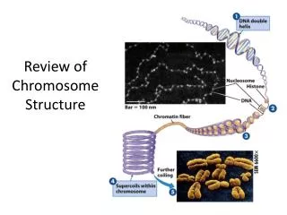
The basic packaging unit of a chromosome in all eukaryotes is the nucleosome. • DNA double strand wounds around eight core histones. Nucleosome consists of 8 core histones and 146 base pairs of DNA. • Neighbouring nucleosomes are connected by the aid of H1, which has a key role in the coiling and packaging of chromatin
The next level is a superhelix formed by nucleosomes and histone H1. This is chromatin. • Chromatin is the combination of DNA and proteins (histones and nonhistones) that makes the chromosome.
All of the cells in a body are somatic cells except from the germ cells (soma,body). • 46 chromosomes in human somatic cells are found as 23 pairs.
22 of these 23 pairs are called as autosomes and they are same in women and men. The biggest one is chromosome 1 and the smallest ones are 21 and 22. They are aligned from the biggest to the smallest.
The other pair is sex chromosomes: XX in females and XY in males.
Chromosome pairs (homolog chromosomes or homologs) carry identical genetic data matching with each other. Members of a pair of chromosomes are known as homologs. They have the same sequencing. • The alternative form of a gene in any specific locus is called as an allele. • One homolog of each chromosome is inherited from mother, and the other one from father.
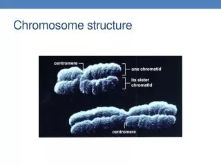
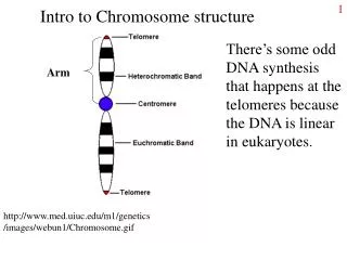
Each chromosome can be seen to consist of two identical strands known as chromatids or sister chromatids, which are the result of DNA replication having taken place during the S (synthesis) phase of the cell cycle. These sister chromatids can be seen to be joined at a primary constriction known as the centromere.
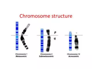
Centromeres consist of several hundred kilobases of repetitive DNA and are responsible for the movement of chromosomes at cell division. • Each centromere divides the chromosome into short and long arms, designated p(petite) and q(‘g’=grande), respectively.
Morphologically chromosomes are classified according to the position of the centromere and this can be distinguished easily in metaphase. These are: _ Metacentric Submetacentric Akrocentric Classification of Chromosomes
MetacentricChromosomes: If centromere is located centrally, the chromosome is metacentric. These chromosomes’ arms are nearly in the same length.
SubmetacentrikChromosomes: If the centromere is in an intermediate position, the chromosome is submetacentric. The length of the arms of the chromosomes are not equal with each other.
AcrocentricChromosomes: If the centromere is located in terminal region, it is acrocentric.
If the centromere is in the end point of the arms and there is only one arm of the chromosome, these are called as telocenric chromosomes. These are not found in normal human karyotype. Sometimes in rearrangements of the chromosomes, they can be seen.
Acrocentrichumanchromosomes (13, 14, 15, 21 and 22.) sometimeshavepartscalledsatellitesthat form thenucleolus of therestinginterphasecellandcontainmultiplerepeatcopies of thegenesforribosomal RNA. Theyareboundedtotheshortarms of acrocentricchromosomesbyseconderconstrictions.
Nomenclature of Chromosomes • Tjio and Levan in 1956 • The number of human chromosomes (2n=46) • The first international meeting in 1960 in Denver • In this meeting a standardization had been accepted for human chromosomes, this nomenclature had been called as Denver System or Classification.
According to Denver system, initially total chromosome number is written, then sex chromosomes and finally chromosomal abnormalities if there is any. _46, XY • 45, X0 • 47,XX,+21 • 46,XY,del(5p)
According to this system, human chromosomes are subdivided into sevengroups (A, B, C, D, E, F, G) and sex chromosomes. The chromosomes are numbered from the biggest to the smallest (1-22) except from sex chromosomes.
There are some basic criteria for the identifying chromosomes: - Their overall length - Centromere position, • The presence of seconder constriction, • Banding characteristics of chromosomes
Karyotype shows each chromosome pair in descending order of size.
The form of stylized ideal karyotype is known as an idiogram.
Group A Chromosomes:Chromosomes numbered as1, 2 and 3. Centromere is located centrally and they are the biggest chromosomes. The biggest chromosome in this group is the first one. Second chomosome’s centromere is a little far away from centromere, so it is called as submedian.
Group B Chromosomes: These are submedian and fourth and fifth chromosomes are in this group. • Group C Chromosomes:These are submedian and from 6th to 12th chromosomes are in this group. • Group D Chromosomes: They are acrocentric chromosomes and they have satellites in their short arms. 13th, 14th and 15th chromosomes are in this group. These are big acrocentric chromomes.
Group E Chromosomes: These are submedian and small.16th, 17th and 18th chromosome are in this group. 16th chromosome is near to median. • Group F Chromosomes: 19th and 20th chromosomes are in this group. These are small and median. They look like butterfly. • Group G Chromosomes: 21st and 22nd chromosomes are in this group. These are small and acrocentric.
Sex Chromosomes:X and Y chromosomes are in this group. They have no number and group. X chromosome resembles to group C and it is near to median. In terms of size, X chromosome is after 6th chromosome. Y chromosome is like the chromosomes in group G. It differs morphologically by staining darkly and the long arms of it are near to each other.
Determination of Chromosomes • In chromosome banding, chromatin, the combination of DNA and histone proteins of which chromosomes are made, exists in two forms. Euchromatin stains lightly and consists of genes that are actively expressed. • In contrast, heterochromatin stains darkly and is made up largely of inactive unexpressed repetitive DNA.
Three different staining methods can be utilized to identify individual chromosomes. • G(Giemsa) banding • Q (Quinacrine) banding • R (Reverse) banding
G banding:This is the method most commonly used. The chromosomes are treated with trypsin, which denaturates their protein content, and then stained with a DNA-binding dye known as Giemsa, which gives each chromosome a characteristic and reproducible pattern of light and dark bands.
Q banding: This gives a banding pattern similar to that obtained with Giemsa, and requires examination of the chromosomes with an ultraviolet fluorescent microscope. Quinacrine is used for this method. • Q banding is useful in determining heteromorphic regions.
R banding:The chromosomes are heat-denaturated before staining with Giemsa, yielding light and dark bands which are the reverse of those obtained using G banding.
Banding Techniques Used in Specific Situations • C (centromeric heterochromatin) banding • High Resolution Banding (HRB) • Fragile Sites
C banding:The centromeres and other heterochromatic regions containing highly repetitive DNA sequences are preferentially stained.
High Resolution Banding (HRB): High resolution banding of the chromosomes at an earlier stage of mitosis, such as prometaphase and prophase, provides greater sensitivity with up to 800 bands per haploid set. • It is especially used when a structural abnormality is suspected.
Fragile Sites:These are the sites that are not stained in characteristic regions of some chromosomes. • It is used especially in Fragile X syndrome. It can be seen in chromosome Xq, near to terminal region.
CLINICAL INDICATIONS FOR CHROMOSOME ANALYSIS • Chromosome analsis is used for some phenotypes in clinical genetics. Some clinical symptomes also require chromosome analysis. These are:
1- Growth and Developmental Problems - - Growth retardation and delay, • Dysmorphic facial appearance, • Multiple malformaton, • Short stature, • ambigious genitalia • Mental retardation
2-Stillbirth and neonatal death 3-Fertility problems 4-Family history 5- Neoplasia 6-Advanced maternal age pregnancies