Download

1 / 25
260 likes | 275 Views
Injuries to the genitourinary tract. *10% of all injuries seen in the emergency room involve the genitourinary system to some extent. *early diagnoses is essential to prevent serious complications. The mnemonic “ABCDE” defines these priorities in order of importance: A, airway

E N D
*10% of all injuries seen in the emergency room involve the genitourinary system to some extent. *early diagnoses is essential to prevent serious complications. The mnemonic “ABCDE” defines these priorities in order of importance: A, airway B, breathing C, circulation D, disability or neurologic status E, exposure (undress)
management of injured pt. should start by. *history include description of the accident, in gunshot wounds, the type & caliber of the weapon should be determined, since high velocity projectile cause much more extensive damage. *physical examination initial assessment should include control of hemorrhage & shock along with resuscitation as required.
after general examination. • examination of abdomen &genitalia for evidence of contusions or subcutaneous hematomas • -Fractures of the lower ribs are often associated with renal injuries • -pelvic fractures often accompany bladder & urethral injuries.
1-catheterization. Blood at the urethral meatus in men indicates urethral injury. So catheterization is contraindicated if blood is present Retrograde urethrography should be done immediately. If no blood is present at the meatus, urethral catheter can be carefully passed to the bladder, microscopic or gross hematuria indicate urinary system injury
2-excretory urography. Immediately after IV line have been established &resuscitation process has began 150ml (2ml/kg) of contrast material can be injected IV. Plain abdominal films permit adequate visualization of the kidneys. It Can be used to detect renal & ureteral injury.
3-retrograde cystography. Filling of the bladder with contrast material is essential to establish whether bladder perforation exist. A film should be obtained with the bladder filled & second after the bladder has emptied itself.
4-urethrography. The urethra will be clearly outlined on film, & extravasation will be visualized if there is injury. 5-Arteiography. may help define renal parenchymal or renal vascular injury.
Injuries to the kidney -Renal injuries are most common injuries of the urinary system. -Fractured rib & transverse vertebral processes may penetrate the renal parenchyma or vasculature.
Etiology. 1-Blunt trauma, (80-85%) of all renal injuries. 2-Pentrating trama Gunshot & knife wounds usually (80%) associated with abdominal visceral injuries 3- deceleration type of injery. Pathology & classification. Pathologic classification of renal injuries is as follow: A-Early pathologic finding Staging of renal injuries.
Grade1 (the most common)—renal contusion or bruising of the renal parenchyma. Subcapsular hematoma without parenchymal laceration. Microscopic hematuria is common, rarely gross hematuria. Grade2—laceration extend (<1cm) in the renal cortex. Perirenal hematoma is usually small. Grade3—the laceration extend through the cortex into the medulla (>1cm)without urinary extravasation. Bleeding can be significant in the presence of large retroperitoneal hematoma. Grade4—the laceration extend into the collecting system. Laceration or thrombosis at a segmental vessel may also present. Grade5—a. multiple major lacerations (shattered kidney). b. avulsion of the renal pedicle or thrombosis of the main renal artery.
B-Late pathologic finding: 1-Urinoma. Deep laceration that are not repaired may result in persistent urinary extravasations. 2-Hydronephrosis. Urinoma compression or heeling by fibrosis can 3-Arteiovenous fistula. 4-Renal vascular hypertension cause stricture and hydronephrosis .
Clinical findings • Symptoms • Hematuria microscopic/gross • Degree does not correlate to severity of injury • In some cases of renal vascular injury there is no hematuria specially in deceleration type • Pain over the flank or all over the abd. • Feature of other abd. Organ injury or shock or peritoneal irritation
signs • Shock and feature of blood loose • Flank echymosis • Lower rib or vertebral fracture • Diffuse abd. Tenderness and acute abd • Large mass representing hematoma or urinoma • Abd. Distension and absent bowel sound • B-Laboratory finding. • *microscopic or gross hematuria. • *normal hematocrit ratio initially later may drop if bleeding persist
C-Staging & x-ray finding. *Abdominal CT is the most direct & effective means of staging renal injuries, it clearly define -the parenchymal laceration -urinary extravasation -retroperitoneal hematoma -identifies the nonviable tissue -outline injuries to the surrounding organs.
Indication for Imaging • Frank hematuria • Microscopic hematuria with shock • Deceleration injury • Evidence of flank trauma • *the major causes of non visualized kidney on IVU • -total pedicle avulsion, • -arterial thrombosis, • -severe contusion causing vascular spasm, • -absence of kidney (congenital or from surgery).
Complications. A-early complications. *Hemorrhage is the most important Pt must be observed closely (monitoring of BP & hematocrit). *Urinary extravasations :prone to abscess formation & sepsis B-late complications -Hypertension, -hydronephrosis, -arteriovenous fistula, -calculus formation, -renal atrophy .
Treatment A .emergency measure prompt treatment of shock & hemorrhage, complete resuscitation, & evaluation of associated injuries. B. surgical measures 1-blunt injuries. 85% of blunt injuries are minor (grade 1&2) & don’t require operation. Bleeding stop spontaneously with bed rest & hydration., antibiotic and vital sign chart
.Indication of surgery • persistent or expanding hematoma • Big or persistent urinary extravasations • Evidence of non viable parenchyma • Renal pedicle injury • Failure of conservative treatment • 2-penetrating injuries • should be surgically explored, except when staging show minor parenchymal injury & no urinary extravasations. • *80% of cases penetrating injuries associated with other organ injuries,
Ureteric injury • Difficult pelvic and abd surgery like hysterectomy • Penetrating injury • Endoscopic injury • C/F • Fever • Flank and lower quadrant pain • Paralytic ileus • Rx by surgery • Principle of repair are • Tension free Speculation • Stinting
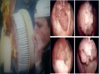
Bladder Injury • Extra peritoneal injury • By direct trauma to empty bladder • Or pelvic fracture • Treated by catheterization • Intera peritoneal injury By direct trauma to full bladder treated by surgery • Both diagnose by cystographyand • clinically have difficult void with hematuria
Urethral Injury • Usually accompany by pelvic fracture • Clinically patient have • Blood in urethral meatus • Penile or perennial ecomosis • Difficult to void • Dx by urethrogram • Rx if no leak of dye that reaching bladder then just cathater If ieak or dye not reach bladder then diveration and late by urethroplasty