Download
1 / 26
260 likes | 420 Views
Veins and lymphatics. By Dr S Homathy. Disorders of veins. Varicose vein Phlebothrombosis Thrombophlebitis. Varicose vein. Abnormally dilated, tortuous veins Produced by prolonged increase in intraluminal pressure and loss of vessel wall support. Types and sites .

E N D
Veins and lymphatics By Dr S Homathy
Disorders of veins • Varicose vein • Phlebothrombosis • Thrombophlebitis
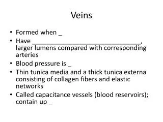
Varicose vein • Abnormally dilated, tortuous veins • Produced by prolonged increase in intraluminal pressure and loss of vessel wall support
Types and sites • Superficial veins of the upper and lower leg • Prolonged standing leads to • Increased venous pressure • venous stasis • Pedal oedema • Females> males • Obesity increases the risk. • Pregnancy • Oesophagealvarices • Haemorrhoids
Clinical features • Home work
Phlebothrombosis and Thrombophlebitis • Deep leg veins acccount of > 90% of cases. • What is Phlebothrombosis (DVT)? • What is Thrombophlebitis ? • What are the predisposing factors / causes of DVT? • Migratory thrombophlebitis?
Superior and inferior vena caval syndrome SVS • Caused by • Neoplasm compress or invade the SVC • Bronchogenic carcinoma / mediastinal lymphoma • Clinical features • Marked dilation of veins of head, neck and arms • Cyanosis • Respiratory distress due to pulmonary vessel compression
IVS • Caused by • Neoplasm compress / invade the vessel (hepatocellular, renal cell carcinoma) • Thrombus from hepatic, renal, lower extremities • Clinical features • Lower extremity oedema • Distension of superficial collateral vein of the lower abdomen • Massive proteinuria in case of renal vein involvement
Lymphangitis and lymphoedema • Causes and consequences? • Home work
Tumours • Benign- haemangioma • Intermediate lesion • Highly malignant – angiosarcoma
Haemangiomas • Very common • Characterized by increased numbers of normal or abnormal vessels filled with blood • Commonly present at birth • Most are expand with growth • Many capillary lesion regress with time
Clinical variant of Haemangiomas Capillary haemangioma • Most common variant • Occurs in the • skin, • subcutaneous tissues • mucous membranes of the oral cavities and lips • Liver • Spleen • kidneys.
Morphology Gross • Bright reed to blue • Few mm to several cm in dm • At the level with surface / slightly elevated • Have intact overlying epithelium Histology • Unencapsulated, aggregates of closely packed, thin – walled capillaries • Usually blood filed and lined by flattened epithelium • Vessels are separated by scant cTstroma • Lumen partioally or completely thrombosed and organised
Cavernous haemangioma • Characterised by large, dilated channels • Less well circumscribed • Frequently involve deep structures.
Morphology Gross • Red- blue, soft, spongy masses Histology • Sharply defined, unencapsulated • Composed of large, cavernous blood - filled vascular spaces separated by a mild to moderate amount of CT stroma • Intravascular thrombosis with associated dystrophic calcification is common
Similar to strawberry hemangiomas but are more deeply situated. • They may appear as a red-blue spongy mass of tissue filled with blood
Pyogenicgranuloma • Is a capillary haemangioma • Rapidly growing peduncular red nodule • Seen on the skin, gingival or oral mucosa • Bleed easily often ulcerated Histology • Proliferating capillaries • Accompanied by extensive oedema • Acute and chronic inflammatory infiltrate