Download
1 / 40
400 likes | 593 Views
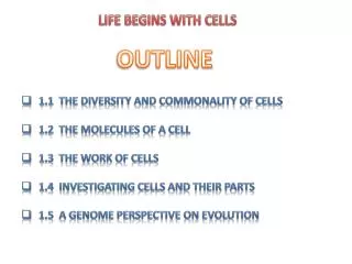
LIFE BEGINS WITH CELLS. OUTLINE. 1.1 The Diversity and Commonality of Cells 1.2 The Molecules of a Cell 1.3 The Work of Cells 1.4 Investigating Cells and Their Parts 1.5 A Genome Perspective on Evolution. 1. 4 Investigating Cells and Their Parts.

E N D
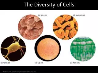
LIFE BEGINS WITH CELLS OUTLINE • 1.1 The Diversity and Commonality of Cells • 1.2 The Molecules of a Cell • 1.3 The Work of Cells • 1.4 Investigating Cells and Their Parts • 1.5 A Genome Perspective on Evolution
1.4 Investigating Cells and Their Parts • To build an integrated understanding of how the various molecular components that underlie cellular functions work together in a living cell, we must draw on various perspectives. • Here, we look at how five disciplines—cell biology, biochemistry, genetics, genomics, and developmental biology—cancontribute to our knowledge of cell structure and function. • The experimental approaches of each field probe the cell’sinner workings in different ways, allowing us to ask different types of questions about cells and what they do. • Cell division provides a good example to illustrate the role ofdifferent perspectives in analyzing a complex cellular process.
The realm of biology ranges in scale more than a billion-fold (Figure 1-20). FIGURE 1-20 Biologists are interested in objects rangingin size from small molecules to the tallest trees.
Beyond that, it’s ecology and earth scienceat the “macro” end, chemistry and physics at the “micro” end. • The visible plants and animals that surround us are measuredin meters (100–102m). • By looking closely, we can see a biological world of millimeters (1 mm = 10-3m) and even tenths of millimeters (10-4m). • Setting aside oddities like chicken eggs, most cells are 1–100 micrometers (1 m = 10-6m) long and thus clearly visible only when magnified. • To see the structures within cells, we must go farther down the size scale to 10–100 nanometers (1 nm = 10-9m).
1.4 Investigating Cells and Their Parts Cell Biology Reveals the Size, Shape,and Location of Cell Components Biochemistry Reveals the Molecular Structureand Chemistry of Purified Cell Constituents Genetics Reveals the Consequencesof Damaged Genes Genomics Reveals Differences in the Structureand Expression of Entire Genomes Developmental Biology Reveals Changes in the Properties of Cells as They Specialize Choosing the Right Experimental Organismfor the Job
Cell Biology Reveals the Size, Shape,and Location of Cell Components • Actual observation of cells awaited development of the first,crude microscopes in the early 1600s. • A compound microscope, the most useful type of light microscope, has twolenses. • The total magnifying power is the product of themagnification by each lens. • As better lenses were invented,the magnifying power and the ability to distinguish closelyspaced objects, the resolution, increased greatly. • Moderncompound microscopes magnify the view about a thousand-fold, so that a bacterium 1 micrometer (1 m) long looks likeit’s a millimeter long. • Objects about 0.2 m apart can be discerned in these instruments.
Microscopy is most powerful when particular components of the cell are stained or labeled specifically, enablingthem to be easily seen and located within the cell. • A simpleexample is staining with dyes that bind specifically to DNAto visualize the chromosomes. • Specific proteins can be detected by harnessing the binding specificity of antibodies,the proteins whose normal task is to help defend animalsagainst infection and foreign substances. • In general, eachtype of antibody binds to one protein or large polysaccharide and no other (Chapter 3). • Purified antibodies can bechemically linked to a fluorescent molecule, which permitstheir detection in a special fluorescence microscope (Chapter 5). • If a cell or tissue is treated with a detergent that partially dissolves cell membranes, fluorescent antibodies candrift in and bind to the specific protein they recognize. • When the sample is viewed in the microscope, the boundfluorescent antibodies identify the location of the target protein (see Figure 1-15).
Better still is pinpointing proteins in living cells with intact membranes. • One way of doing this is to introduce anengineered gene that codes for a hybrid protein: part of thehybrid protein is the cellular protein of interest; the otherpart is a protein that fluoresces when struck by ultravioletlight. • A common fluorescent protein used for this purposeis green fluorescent protein (GFP), a natural protein thatmakes some jellyfish colorful and fluorescent. • GFP “tagging” could reveal, for instance, that a particular proteinis first made on the endoplasmic reticulum and then ismoved by the cell into the lysosomes. • In this case, first theendoplasmic reticulum and later the lysosomes would glowin the dark.
Chromosomes are visible in the light microscope onlyduring mitosis, when they become highly condensed. • The extraordinary behavior of chromosomes during mitosis firstwas discovered using the improved compound microscopesof the late 1800s. • About halfway through mitosis, the replicated chromosomes begin to move apart. • Microtubules, oneof the three types of cytoskeletal filaments, participate in thismovement of chromosomes during mitosis. • Fluorescent tagging of tubulin, the protein subunit that polymerizes to formmicrotubules, reveals structural details of cell division thatotherwise could not be seen and allows observation of chromosome movement (Figure 1-21).
FIGURE 1-21 During the later stages of mitosis,microtubules (red) pull the replicated chromosomes (black)toward the ends of a dividing cell.
Electron microscopes use a focused beam of electrons instead of a beam of light. • In transmission electron microscopy,specimens are cut into very thin sections and placed under ahigh vacuum, precluding examination of living cells. • The resolution of transmission electron microscopes, about 0.1 nm,permits fine structural details to be distinguished, and theirpowerful magnification would make a 1-m-long bacterialcell look like a soccer ball. • Most of the organelles in eukaryotic cells and the double-layered structure of the plasmamembrane were first observed with electron microscopes(Chapter 5). • With new specialized electron microscopy techniques, three-dimensional models of organelles and largeprotein complexes can be constructed from multiple images. • But to obtain a more detailed look at the individual macromolecules within cells, we must turn to techniques within thepurview of biochemistry.
Biochemistry Reveals the Molecular Structure and Chemistry of Purified Cell Constituents • Biochemists extract the contents of cells and separate theconstituents based on differences in their chemical or physical properties, a process called fractionation. • Of particularinterest are proteins, the workhorses of many cellularprocesses. • A typical fractionation scheme involves use ofvarious separation techniques in a sequential fashion. • These separation techniques commonly are based on differences in the size of molecules or the electrical charge ontheir surface (Chapter 3). • To purify a particular protein ofinterest, a purification scheme is designed so that each stepyields a preparation with fewer and fewer contaminatingproteins, until finally only the protein of interest remains(Figure 1-22).
FIGURE 1-22 Biochemical purification of a protein from acell extract often requires several separation techniques.
The initial purification of a protein of interest from a cellextract often is a tedious, time-consuming task. • Once a smallamount of purified protein is obtained, antibodies to it canbe produced by methods discussed in Chapter 6. • For a biochemist, antibodies are near-perfect tools for isolating largeramounts of a protein of interest for further analysis. • In effect,antibodies can “pluck out” the protein they specifically recognize and bind from a semipure sample containing numerous different proteins. • An increasingly common alternative isto engineer a gene that encodes a protein of interest with asmall attached protein “tag,” which can be used to pull outthe protein from whole cell extracts.
Purification of a protein is a necessary prelude to studieson how it catalyzes a chemical reaction or carries out otherfunctions and how its activity is regulated. • Some enzymes aremade of multiple protein chains (subunits) with one chaincatalyzing a chemical reaction and other chains regulatingwhen and where that reaction occurs. • The molecular machines that perform many critical cell processes constituteeven larger assemblies of proteins. • By separating the individual proteins composing such assemblies, their individual catalytic or other activities can be assessed. • For example,purification and study of the activity of the individual proteins composing the DNA replication machine providedclues about how they work together to replicate DNA duringcell division (Chapter 4).
The folded, three-dimensional structure, or conformation, of a protein is vital to its function. • To understand the relation between the function of a protein and its form, weneed to know both what it does and its detailed structure. • The most widely used method for determining the complexstructures of proteins, DNA, and RNA is x-ray crystallography. • Computer-assisted analysis of the data often permits thelocation of every atom in a large, complex molecule to be determined. • The double-helix structure of DNA, which is keyto its role in heredity, was first proposed based on x-ray crystallographic studies. • Throughout this book you will encounter numerous examples of protein structures as we zeroin on how proteins work.
Genetics Reveals the Consequencesof Damaged Genes • Biochemical and crystallographic studies can tell us muchabout an individual protein, but they cannot prove that it isrequired for cell division or any other cell process. • The importance of a protein is demonstrated most firmly if a mutation that prevents its synthesis or makes it nonfunctionaladversely affects the process under study.
We define the genotype of an organism as its compositionof genes; the term also is commonly used in reference to different versions of a single gene or a small number of genesof interest in an individual organism. • A diploid organismgenerally carries two versions (alleles) of each gene, one derived from each parent. • There are important exceptions, suchas the genes on the X and Y chromosomes in males of somespecies including our own. • The phenotype is the visible outcome of a gene’s action, like blue eyes versus brown eyes orthe shapes of peas. • In the early days of genetics, the locationand chemical identity of genes were unknown; all that couldbe followed were the observable characteristics, the phenotypes. • The concept that genes are like “beads” on a long“string,” the chromosome, was proposed early in the 1900sbased on genetic work with the fruit fly Drosophila.
In the classical genetics approach, mutants are isolatedthat lack the ability to do something a normal organism cando. • Often large genetic “screens” are done, looking for manydifferent mutant individuals (e.g., fruit flies, yeast cells) thatare unable to complete a certain process, such as cell divisionor muscle formation. • In experimental organisms or culturedcells, mutations usually are produced by treatment with amutagen, a chemical or physical agent that promotes mutations in a largely random fashion. • But how can we isolateand maintain mutant organisms or cells that are defective insome process, such as cell division, that is necessary for survival?
One way is to look for temperature-sensitive mutants. • These mutants are able to grow at one temperature, the permissive temperature, but not at another, usually higher temperature, the nonpermissive temperature. • Normal cells cangrow at either temperature. • In most cases, a temperature-sensitive mutant produces an altered protein that works atthe permissive temperature but unfolds and is nonfunctionalat the nonpermissive temperature. • Temperature-sensitivescreens are readily done with viruses, bacteria, yeast, round-worms, and fruit flies.
By analyzing the effects of numerous different temperature-sensitive mutations that altered cell division, geneticists discovered all the genes necessary for cell division without knowinganything, initially, about which proteins they encode or howthese proteins participate in the process. • The great power of genetics is to reveal the existence and relevance of proteins without prior knowledge of their biochemical identity or molecularfunction. • Eventually these “mutation-defined” genes were isolated and replicated (cloned) with recombinant DNA techniques discussed in Chapter 9. • With the isolated genes in hand,the encoded proteins could be produced in the test tube or inengineered bacteria or cultured cells. • Then the biochemistscould investigate whether the proteins associate with other proteins or DNA or catalyze particular chemical reactions duringcell division (Chapter 21).
The analysis of genome sequences from various organisms during the past decade has identified many previouslyunknown DNA regions that are likely to encode proteins(i.e., protein-coding genes). • The general function of the protein encoded by a sequence-identified gene may be deducedby analogy with known proteins of similar sequence. • Ratherthan randomly isolating mutations in novel genes, severaltechniques are now available for inactivating specific genesby engineering mutations into them (Chapter 9). • The effectsof such deliberate gene-specific mutations provide information about the role of the encoded proteins in living organisms. • This application of genetic techniques starts with agene/protein sequence and ends up with a mutant phenotype;traditional genetics starts with a mutant phenotype and endsup with a gene/protein sequence.
Genomics Reveals Differences in the Structure and Expression of Entire Genomes • Biochemistry and genetics generally focus on one gene and itsencoded protein at a time. • While powerful, these traditionalapproaches do not give a comprehensive view of the structure and activity of an organism’s genome, its entire set ofgenes. • The field of genomics does just that, encompassing themolecular characterization of whole genomes and the determination of global patterns of gene expression. • The recentcompletion of the genome sequences for more than 80species of bacteria and several eukaryotes now permits comparisons of entire genomes from different species. • The resultsprovide overwhelming evidence of the molecular unity of lifeand the evolutionary processes that made us what we are (seeSection 1.5). • Genomics-based methods for comparing thousands of pieces of DNA from different individuals all at thesame time are proving useful in tracing the history and migrations of plants and animals and in following the inheritance of diseases in human families.
New methods using DNA microarrays can simultaneously detect all the mRNAs present in a cell, thereby indicating which genes are being transcribed. • Such globalpatterns of gene expression clearly show that liver cells transcribe a quite different set of genes than do white blood cellsor skin cells. • Changes in gene expression also can be monitored during a disease process, in response to drugs or otherexternal signals, and during development. • For instance, therecent identification of all the mRNAs present in cultured fibroblasts before, during, and after they divide has given usan overall view of transcriptional changes that occur duringcell division (Figure 1-23). • Cancer diagnosis is being transformed because previously indistinguishable cancer cellshave distinct gene expression patterns and prognoses (Chapter 23). • Similar studies with different organisms and celltypes are revealing what is universal about the genes involvedin cell division and what is specific to particular organisms.
FIGURE 1-23 DNA microarray analysis gives a globalview of changes in transcription following addition of serumto cultured human cells.
The entire complement of proteins in a cell, its proteome,is controlled in part by changes in gene transcription. • Theregulated synthesis, processing, localization, and degradationof specific proteins also play roles in determining the proteome of a particular cell, and the association of certain proteins with one another is critical to the functional abilitiesof cells. • New techniques for monitoring the presence and interactions of numerous proteins simultaneously, called proteomics, are one way of assembling a comprehensive viewof the proteins and molecular machines important for cellfunctioning. • The field of proteomics will advance dramatically once high-throughput x-ray crystallography, currentlyunder development, permits researchers to rapidly determinethe structures of hundreds or thousands of proteins.
Developmental Biology Reveals Changes in the Properties of Cells as They Specialize • Another approach to viewing cells comes from studying howthey change during development of a complex organism. • Bacteria, algae, and unicellular eukaryotes (protozoans,yeasts) often, but by no means always, can work solo. • Theconcerted actions of the trillions of cells that compose ourbodies require an enormous amount of communication anddivision of labor. • During the development of multicellular organisms, differentiation processes form hundreds of celltypes, each specialized for a particular task: transmission ofelectrical signals by neurons, transport of oxygen by redblood cells, destruction of infecting bacteria by macrophages, contraction by muscle cells, chemical processing byliver cells.
Many of the differences among differentiated cells aredue to production of specific sets of proteins needed to carryout the unique functions of each cell type. • That is, only asubset of an organism’s genes is transcribed at any given timeor in any given cell. • Such differential gene expression at different times or in different cell types occurs in bacteria, fungi,plants, animals, and even viruses. • Differential gene expression is readily apparent in an early fly embryo in which allthe cells look alike until they are stained to detect the proteins encoded by particular genes (Figure 1-24). • Transcription can change within one cell type in response to anexternal signal or in accordance with a biological clock;some genes, for instance, undergo a daily cycle between lowand high transcription rates.
FIGURE 1-24 Differential gene expression can bedetected in early fly embryos before cells aremorphologically different.
Producing different kinds of cells is not enough to make anorganism, any more than collecting all the parts of a truck inone pile gives you a truck. • The various cell types must be organized and assembled into all the tissues and organs. • Evenmore remarkable, these body parts must work almost immediately after their formation and continue working during thegrowth process. • For instance, the human heart begins to beatwhen it is less than 3 mm long, when we are mere 23-day-oldembryos, and continues beating as it grows into a fist-sizemuscle. • From a few hundred cells to billions, and still ticking.
In the developing organism, cells grow and divide atsome times and not others, they assemble and communicate,they prevent or repair errors in the developmental process,and they coordinate each tissue with others. • In the adult organism, cell division largely stops in most organs. • If part ofan organ such as the liver is damaged or removed, cell division resumes until the organ is regenerated. • The legend goesthat Zeus punished Prometheus for giving humans fire bychaining him to a rock and having an eagle eat his liver. • Thepunishment was eternal because, as the Greeks evidentlyknew, the liver regenerates.
Developmental studies involve watching where, when,and how different kinds of cells form, discovering which signals trigger and coordinate developmental events, and understanding the differential gene action that underliesdifferentiation (Chapters 15 and 22). • During developmentwe can see cells change in their normal context of other cells. • Cell biology, biochemistry, cell biology, genetics, and genomics approaches are all employed in studying cells duringdevelopment.
Choosing the Right Experimental Organismfor the Job • Our current understanding of the molecular functioning ofcells rests on studies with viruses, bacteria, yeast, protozoa,slime molds, plants, frogs, sea urchins, worms, insects, fish,chickens, mice, and humans. • For various reasons, some organisms are more appropriate than others for answering particular questions. • Because of the evolutionary conservationof genes, proteins, organelles, cell types, and so forth, discoveries about biological structures and functions obtainedwith one experimental organism often apply to others. • Thusresearchers generally conduct studies with the organism thatis most suitable for rapidly and completely answering thequestion being posed, knowing that the results obtained inone organism are likely to be broadly applicable. • Figure 1-25summarizes the typical experimental uses of various organisms whose genomes have been sequenced completely ornearly so. • The availability of the genome sequences for theseorganisms makes them particularly useful for genetics andgenomics studies.
FIGURE 1-25 Each experimental organism used in cellbiology has advantages for certain types of studies.
Bacteria have several advantages as experimental organisms: • They grow rapidly, possess elegant mechanisms forcontrolling gene activity, and have powerful genetics. • Thislatter property relates to the small size of bacterial genomes,the ease of obtaining mutants, the availability of techniquesfor transferring genes into bacteria, an enormous wealth ofknowledge about bacterial gene control and protein functions, and the relative simplicity of mapping genes relativeto one another in the genome. • Single-celled yeasts not onlyhave some of the same advantages as bacteria, but also possess the cell organization, marked by the presence of a nucleus and organelles, that is characteristic of all eukaryotes.
Studies of cells in specialized tissues make use of animaland plant “models,” that is, experimental organisms with attributes typical of many others. • Nerve cells and muscle cells,for instance, traditionally were studied in mammals or increatures with especially large or accessible cells, such as thegiant neural cells of the squid and sea hare or the flight muscles of birds. • More recently, muscle and nerve developmenthave been extensively studied in fruit flies (Drosophilamelanogaster), roundworms (Caenorhabditiselegans), andzebrafish in which mutants can be readily isolated. • Organisms with large-celled embryos that develop outside themother (e.g., frogs, sea urchins, fish, and chickens) are extremely useful for tracing the fates of cells as they form different tissues and for making extracts for biochemical studies. • Forinstance, a key protein in regulating mitosis was firstidentified in studies with frog and sea urchin embryosand subsequently purified from extracts (Chapter 21).
Using recombinant DNA techniques researchers can engineer specific genes to contain mutations that inactivateor increase production of their encoded proteins. • Suchgenes can be introduced into the embryos of worms, flies,frogs, sea urchins, chickens, mice, a variety of plants, andother organisms, permitting the effects of activating a geneabnormally or inhibiting a normal gene function to be assessed. • This approach is being used extensively to producemouse versions of human genetic diseases. • New techniquesspecifically for inactivating particular genes by injectingshort pieces of RNA are making quick tests of gene functions possible in many organisms.
Mice have one enormous advantage over other experimental organisms: they are the closest to humans of any animal for which powerful genetic approaches are feasible. • Engineered mouse genes carrying mutations similar to thoseassociated with a particular inherited disease in humans canbe introduced into mouse embryonic stem (ES) cells. • Thesecells can be injected into an early embryo, which is then implanted into a pseudopregnant female mouse (Chapter 9). • Ifthe mice that develop from the injected ES cells exhibit diseases similar to the human disease, then the link between thedisease and mutations in a particular gene or genes is supported. • Once mouse models of a human disease are available, further studies on the molecular defects causing thedisease can be done and new treatments can be tested,thereby minimizing human exposure to untested treatments.
A continuous unplanned genetic screen has been performed on human populations for millennia. • Thousands ofinherited traits have been identified and, more recently,mapped to locations on the chromosomes. • Some of thesetraits are inherited propensities to get a disease; others areeye color or other minor characteristics. • Genetic variations invirtually every aspect of cell biology can be found in humanpopulations, allowing studies of normal and disease statesand of variant cells in culture.
Less-common experimental organisms offer possibilitiesfor exploring unique or exotic properties of cells and forstudying standard properties of cells that are exaggerated ina useful fashion in a particular animal. • For example, the endsof chromosomes, the telomeres, are extremely dilute in mostcells. • Human cells typically contain 92 telomeres (46 chromosomes 2 ends per chromosome). • In contrast, some protozoa with unusual “fragmented” chromosomes containmillions of telomeres per cell. • Recent discoveries abouttelomere structure have benefited greatly from using this natural variation for experimental advantage.