Download
1 / 38
380 likes | 489 Views
Muscle system. Characteristics of Muscles. Irritability (excitability) Respond to chemicals (neurotransmitters), stretch etc. by altering their plasma membrane electrical charge Contractility Can shorten their length by a substantial amount when stimulated Extensibility

E N D
Characteristics of Muscles • Irritability (excitability) • Respond to chemicals (neurotransmitters), stretch etc. by altering their plasma membrane electrical charge • Contractility • Can shorten their length by a substantial amount when stimulated • Extensibility • Can stretch back to their original length after each contraction • Elasticity • When stretched and then released, muscle will recoil to its original resting length
Muscle Functions • Movement • Body movement and mobility and various bodily functions • Posture and body support • Maintain body position • Heat production • 85% body heat is generated by muscle contractions
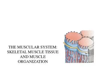
Muscle types • Skeletal muscle • Voluntary • Striated • Usually attached to bone • Smooth muscle • “fusiform” cells • Involuntary • “Pacemaker” cells usually spontaneously depolarize and stimulate contractions (autorhythmic) • Autonomic nervous system can stimulate contractions as well • Cardiac muscle (heart muscle) • Involuntary • “pacemaker” cells autorhythmic • Autonomic nervous system control • Striated, like skeletal muscle cells • Remember the intercalated discs?
Skeletal muscle - Attachments • Usually attached to 2 or more bones (span a joint) • Voluntary: under voluntary/conscious control • Skeletal muscle cells = “muscle fibers” or “myofibers” • Range from 3-30 cm long • Attached to the bone by tendons (in most cases)
Skeletal muscle – Attachments • “sheaths” around muscle fibers • Endomysium: areolar connective tissue • Where blood capillaries, neural inputs are located • Perimysium: bundles a number of muscle cells together • The bundles of muscle cells = “fascicles” • Epimysium: surrounds the entire muscle • Deep fasciae: separates adjacent muscles • Devoid of adipose cells. • Superficial fasciae (hypodermis): between muscle and dermis (skin) • Very adipose-cell dense in some areas (abdomen & buttocks • Note: The structures attaching the muscle are extension of the connective tissue forming the endo/peri/epimysium
Muscle structures - Attachments • Deep fascia: fibrous connective tissue anchoring groups of muscle and deep structures, support nerves, blood vessels, forms tendons, ligaments, joint capsules. • Superficial fasciae: support the skin, nerves and blood vessels, provides elasticity to hypodermis • Aponeuroses: sheet-like tendons- ex: over the skull • Tendon sheath: encloses group of thin tendon in a sheath, for protection and lubrication– wrist. • Retinaculum:sheat of connective tissue binding a group of tendons
Aponeuroses in scalp and abdomen Retinaculum in your wrist
Applications • Compartment syndrome - due to muscle swelling In order to prevent limb loss, the tissues are slashed
Muscle attachment • Epimysium: surrounds the entire muscle • Recall that tendons usually attach to bone via the periosteum (outer membrane on a bone) • 2 types of muscle-to-bone connections: • “direct” or “fleshy attachment” • Collagen fibers of epimysium continue in the periosteum • Looks like the muscle is directly attached to bone (in your ribs) • “indirect” attachment • Epimysium is in unison with tendon • The tendon attaches to bone • Seen as a white or pale “gap” between the red part of a muscle and the bone
Skeletal muscle cells • Derived from stem cells called “myoblasts” • Several myoblasts will fuse together during embryonic development • Reason why skeletal muscle cells are multinucleate (have many nuclei)
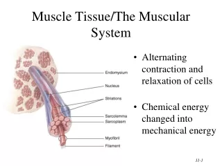
Skeletal muscle cells • Muscle cell (myocyte, muscle fiber) anatomy: • Outer plasma membrane = sarcolemma (“flesh-lining”) • Sarcoplasm: cytoplasm of the myocyte • Myofibrils: Bundles of protein running along the length of the cell • Myoglobin: similar to hemoglobin • Stores oxygen within the muscle cell • Important for ATP synthesis during contraction
Skeletal muscle cells • Muscle cell (myocyte, muscle fiber) anatomy: • Sarcoplasmic reticulum: specialized area of the endoplasmic reticulum • Surrounds the myofibrils like a web • At each end, forms bag-like structure “terminal cisternae” • Area where calcium (Ca2+) is stored • Transverse tubules: tube formation of the sarcolemma • Tubes run from the external surface straight through the muscle cell • Transverse tubules and terminal cisternae form a “triad” structure (many of these along the length of the muscle cell)
Transverse tubules form a “pore” through which the muscle cell can “sense” it’s outside environment and respond to stimuli
Myofibrils • Made of “myofilaments”: • Thick myofilaments: bundles of myosin • Thin myofilament: Actin
Myosin • Myosin head on the thick filament will try to bind to the actin bead at a specific “active” site on the actin molecule • Also has “control” proteins to control contraction (control myosin attachment)
Actin • Thin myofilaments: • Globular actin filamentous actin • Tropomyosin: “plugs” the site where myosin wants to attach • Troponin: calcium-sensitive protein
The sarcomere • “sarcomere”: refers to the entire complex between each Z-disc. • Formation of bands: • - A bands: Myosin + actin myofilaments • - I band: actin only
In summary http://www.life.uiuc.edu/crofts/bioph354/lect16&17.html
Motor unit • All muscle fibers controlled by 1the same motor neuron • Which of the two set up provides the finest muscle control? • 10 muscle fibers controlled by 1 motor neuron • Or • 1000 muscle fibers controlled by 1 motor neuron
Contraction • During contraction, the myosin “rows” along the actin and pull the Z-discs closer to one another • The I band shortens • The A band do not change • http://www.blackwellpublishing.com/matthews/myosin.html • http://www.ebsa.org/npbsn41/intro_muscle.html
Skeletal muscle terms • “origin” = area where muscle attaches to bone that does not move (remains stationary) • “belly” = thick region of the muscle • “insertion” = area where muscle attaches to bone that moves
Skeletal muscle anatomy • Different forms of skeletal muscle: • Fusiform: thick in the middle, thin at ends • Biceps branchii, gastrocnemius in calf • Parallel: long, strap-like • Spans great lengths, can shorten/contract more than other muscles • Weaker than fusiform muscles • Rectus abdominus (abs…6-pack) • Convergent: fan shaped/leaf shaped • Large middle that tapers or focuses on 1 narrow insertion • Pectoralis major in the chest • Pennate and circular muscles (next slide)
Skeletal muscle anatomy • Pennate muscles: feather shaped - The muscle fibers attach obliquely or at an angle to the tendon • Unipennate: all muscles approach on 1 side of the tendon • Palmar interosseus of the hand • Bipennate: muscle fibers attach on both sides of the tendon • Rectus femorus of the thigh • Multipennate: a branched tendon, with muscle fibers attaching throughout • Deltoid of the shoulder • Circular muscle: sphincters etc. - Forms a ring around an opening/passage • Orbicularis oculi around the eye • Anal sphincter
Skeletal muscle anatomy • Each muscle requires nutrients & oxygen • Each muscle usually has at least 1 artery entering • Larger muscles (quadriceps femoris etc.) have 2+ arteries to bring blood etc. • Contraction requires a great deal of skeletal muscle cell metabolic activity
Skeletal muscle terms • Insertion & origin will differ depending on the MOTION • Quadriceps muscle: • When you kick (extend your knee), the quadriceps moves the tibia • Tibia is the insertion (moves), and the femur is the origin (stays still) • When you sit down, the terminology changes: • Tibia stays still (origin), and the femur moves (insertion)
Skeletal muscle terminology • Common muscle terms: • Shape terms (rhomboid, trapezius) • Location (pectoralis, intercostal, brachii, femoris) • Attachment (zygomaticus, temporalis, sternocleidomastoid) • Size (maximus, medius, minimus, longus, brevis) • Fiber orientation • Rectus = straight fibers (rectus abdominus) • Transverse = across (transverse abdominal muscle) • Oblique = slant/angle (external oblique muscle) • Relative position (lateral, medial, internal, external) • Function (adductor, abductor, extensor, flexor, pronator, levator)
Skeletal muscle movements • “action” = movement produced by a muscle • Note that skeletal muscle rarely acts independently or alone…almost always acts in groups • 4 major muscle action categories: • Prime mover (agonist) • Muscle that produces the most force during a particular joint movement • When you flex your elbow, your prime mover is the biceps brachii
Skeletal muscle movements • 4 major muscle action categories: • Prime mover (agonist) • Synergist • Muscle (s) that aid the prime mover • Usually act to stabilize the joint…focus the energy of the prime mover efficiently • Antagonist • Opposes the prime mover (muscles usually act in pairs…prime mover and antagonist) • Fixator • Prevents inappropriate movement • When you flex your elbow, you use the biceps brachii
Diseases of myofilaments • Muscular dystrophy: mutations in the dystrophin gene • Different types: • Duchenne dystrophy • Becker dystrophy
Myasthenia gravis Autoimmune disease: antibodies destroy the Acetylcholine receptors muscle weakness
Tetanus Due to a bacterial toxin http://medinfo.ufl.edu/year2/mmid/bms5300/images/a5.jpg http://www.humanillnesses.com/original/images/hdc_0001_0003_0_img0264.jpg www.4to40.com/images/qa/tetanus.jpg
Types of muscle contractions • Isometric contraction: contraction without a change in muscle length • When you hold in one place…it takes muscles to hold your book in front of you, even if none of the muscles in your arm are changing length • Isotonic contraction: contraction resulting in a change in muscle length, but no alteration in muscle tension • When you increase the internal tension in the muscle to overcome the resistance
Aging and muscle • Varies with people • Decrease in muscle mass • Very dependent on the person’s level of exercise • “If you don’t use it, you loose it”
Muscle imbalances • Eye muscles: various diseases, ex: strabismus • Torticolis