Download

1 / 48
630 likes | 1.31k Views
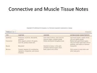
Histology 2 – Connective Tissue. Scott.lehbauer@lethbridgecollege.ab.ca. Connective Tissue Facts. They are the most abundant and widely distributed tissue type in the body.

E N D
Histology 2 – Connective Tissue Scott.lehbauer@lethbridgecollege.ab.ca
Connective Tissue Facts • They are the most abundant and widely distributed tissue type in the body. • Connective tissues run the gamut for vascularity. Some tissues are avascular (Cartilage), some are poorly vascularized (dense connective tissue), and some have rich blood supplies (bone).
Connective Tissue Facts • Connective tissues can be rigid (bone), flexible (adipose), or fluid (blood). • Unlike the tightly packed Epithelial tissues, living cells in connective tissues are separated by a non-living extracellular matrix (Ground Substance and Fibers). • Due to the matrix, connective tissues are able to bear weight, withstand tension, and endure abuses that no other tissues could tolerate.
Connective Tissue Facts • Connective Tissues have many specific functions. Its major functions include • Binding and support • Protection • Insulation • Transportation of substances
Connective Tissue Facts • Connective Tissues are made of three main components: • Ground Substance • Fibers • Cells
Connective Tissue Facts – Ground Substance • The ground substance is the unstructured material between cells that contains the fibers. • The ground substance holds large amounts of fluid and serves as a medium through which nutrients and other substances can diffuse between blood vessels and the cells.
Ground Substance Ground Substance
Connective Tissue Facts - Fibers • There are three types of fibers prevalent in Connective tissues • A.) collagen fibers – are wide and wavy in appearance and generally stain pink. 79% of the protein in the body is collagen. • B.) elastic fibers – are thin flexible fibers made from the protein elastin, that generally stain black. • C.) reticular fibers – are actually thin collagen fibers. They have a spider web appearance and appear black under stain.
Fiber Types Elastic Fiber Reticular Fibers Collagen Fiber
Connective Tissue Facts – The Cells • Each major type of connective tissue has its own fundamental cell type in both immature and mature forms
Cell Types Type of Connective Tissue Immature Cell Mature Cell Connective Tissue Proper Fibroblast Fibrocyte Cartilage Chondroblast Chondrocyte Bone Osteoblast Osteocyte Blood Hematopoietic Stem Cell Blood cell (macrophages)
Other Cells Present • Connective tissue is also home to many other cell types including Fat Cells, and mobile cells that migrate into the connective tissue from the blood stream, ie. mast cells and macrophages.
Connective Tissue Proper - Areolar Connective Tissue Structure- • gel like matrix with all three fiber types present. • Three cell types present • Mast cells • Macrophages • fibroblasts
Connective Tissue Proper - Areolar Connective Tissue • Mast Cells – produce heparin and histamine. • Macrophages - are “big eaters”. They eat bacteria and dead or dying cells. • Fibroblasts–fiber builders.
Connective Tissue Proper - Areolar Connective Tissue Fibroblast Location – • Found between the skin and muscle. • Also found between muscles • Packages organs • Surrounds Capillaries Elastic Fiber Collagen Fiber
Connective Tissue Proper - Areolar Connective Tissue Function – • Wraps and cushions organs. • Macrophages phagocytize bacteria • Holds and conveys tissue fluid.
Connective Tissue Proper - Areolar Connective Tissue • Areolar Connective Tissue is the most widely distributed connective tissue in the body. It serves as a kind of packaging material between other tissues.
Connective Tissue Proper – Adipose Tissue Nucleus Structure – • Adipocytes(fat filled cells) are ring shaped cells filled with tryglycerides. • Has a chicken wire appearance. Adipocyte
Connective Tissue Proper – Adipose Tissue Location – • Found around organs, joints, surrounding the eyeball, within the abdomen.
Connective Tissue Proper – Adipose Tissue Function – • Shock absorption • Energy Storage • Protection • Insulation
Connective Tissue Proper – Reticular Tissue Structure – • A network of reticular fibers with macrophages interspersed.
Connective Tissue Proper – Reticular Tissue Reticular Fibers Location – • Spleen • Lymph nodes • liver Macrophage
Connective Tissue Proper – Reticular Tissue Function • In Lymph Nodes – macrophages devour bacteria, viruses and cancer cells. • In Spleen – macrophages break down dying RBC’s. • In Liver – macrophages (Kupffer cells) devour bacteria. • This tissue forms a soft internal skeleton that supports other cell types.
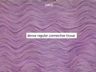
Connective Tissue Proper – Dense Regular Connective Tissue Structure – • Parallel collagen fibers. • Dark colored fibroblasts interspersed.
Connective Tissue Proper – Dense Regular Connective Tissue Collagen Fiber Location – • In tendons and ligaments. • In scar tissue • aponeuroses Fibroblast
Connective Tissue Proper – Dense Regular Connective Tissue Function – • Provide high tensile strength in one direction. • Attached muscles to bone, bone to bone
Hyaline Cartilage Perichondrium Structure – • The chondrocytes (cells) lie in lacunae (spaces around cells). • There is a large amount of extracellular matrix which is bordered on either side by the perichondrium. Chondrocyte Lacunae
Hyaline Cartilage Location - • Forms most of the embryonic skeleton. • Covers the ends of long bones. • Costal cartilage • Nose • Trachea • Larynx
Hyaline Cartilage Extracellular matrix Function – • Provides a smooth surface for joints to move over. • Resists compression and provides flexible support. Lacunae Chondrocyte
Elastic Cartilage Structure- • Similar to Hyaline cartilage with the exception of the elastic fibers in the matrix.
Elastic Cartilage Location – • Ears • Epiglottis Elastic Fiber
Elastic Cartilage Function – • Maintains shape of a structure but is also extremely flexible. Lacunae Chondrocyte
Fibrocartilage Structure – • Thick collagen fibers predominate the matrix. • Chondrocytes are interspersed among the fibers.
Fibrocartilage Location – • Intervertebral Discs • Pubic Symphysis • Menisci of the knee Chondrocyte Collagen Fiber
Fibrocartilage Function – • Shock absorption • Smooth tough support Chondrocyte Collagen Fiber
Compact Bone Osteocyte in Lacunae Structure – • Bone is highly vascular • Haversian canals contain an artery vein and nerve • Osteocytes reside in lacunae within the matrix • Consists of a hard calcified matrix • Canaliculi branch away from the haversian canal to take nutrients to the osteocytes Canaliculi Haversian Canal
Compact Bone Location – • in compact bone • The Haversian system is only found in compact bone • Spongy bone has osteoblasts and osteocytes but no Haversian systems
Compact Bone Lamella Function – • Provides stability to the body and allows for movement with attachment of muscles.
Blood Leukocyte (Neutrophil) Thrombocyte Structure – • Red (Erythrocytes) and White (Leukocytes) blood cells. • There are also platelets (Thrombocytes) • Cells are immersed in a fluid matrix (plasma) Erythrocyte
Blood Location – • Contained within blood vessels and heart.
Blood Function – • Transport of gases • Transport of nutrients • Transport of waste
Practice Quiz • 1.) Name this type of Connective Tissue.
Practice Quiz 2.) a.) Name the structure labeled A. b.) Name the structure labeled B. A B
Practice Quiz 3.) Name one function of this tissue?
Practice Quiz 4.) Where in the body would you find this tissue?
Practice Quiz 5.) Name the fiber type labeled A. A
Answers 1.) Reticular Tissue 2.) a.) osteocyte b.) Haversian Canal 3.) Shock absorption, Insulation, Protection, Energy Storage 4.) Intervertebral Discs, Pubic symphysis, menisci of the knee 5.) Elastic Fiber