Download
1 / 32
320 likes | 399 Views
Cell Structure and Function. Chapter 4. Early Discoveries. Mid 1600s - Robert Hooke observed and described cells in cork Late 1600s - Antony van Leeuwenhoek observed sperm, microorganisms 1820s - Robert Brown observed and named nucleus in plant cells. Developing Cell Theory.

E N D
Cell Structure and Function Chapter 4
Early Discoveries • Mid 1600s - Robert Hooke observed and described cells in cork • Late 1600s - Antony van Leeuwenhoek observed sperm, microorganisms • 1820s - Robert Brown observed and named nucleus in plant cells
Developing Cell Theory • Matthias Schleiden • Theodor Schwann • Rudolf Virchow
Cell Theory 1) Every organism is composed of one or more cells 2) Cell is smallest unit having properties of life 3) Continuity of life arises from growth and division of single cells
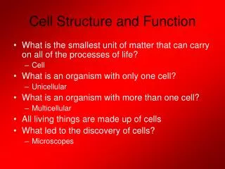
Cell • Smallest unit of life • Can survive on its own or has potential to do so • Is highly organized for metabolism • Senses and responds to environment • Has potential to reproduce
Structure of Cells All start out life with: • Plasma membrane • Region where DNA is stored • Cytoplasm Two types: • Prokaryotic • Eukaryotic
one layer of lipids one layer of lipids Lipid Bilayer • Main component of cell membranes • Gives the membrane its fluid properties • Two layers of phospholipids Figure 4.3Page 56
Membrane Proteins Recognition protein Receptor protein extracellular environment lipid bilayer cytoplasm Protein pump across bilayer Protein channel across bilayer Protein pump Figure 4.4Page 57
Why Are Cells So Small? • Surface-to-volume ratio • The bigger a cell is, the less surface area there is per unit volume • Above a certain size, material cannot be moved in or out of cell fast enough
Microscopes • Create detailed images of something that is otherwise too small to see • Light microscopes • Simple or compound • Electron microscopes • Transmission EM or Scanning EM
Eukaryotic Cells • Have a nucleus and other organelles • Eukaryotic organisms • Plants • Animals • Protistans • Fungi
Animal Cell Features • Plasma membrane • Nucleus • Ribosomes • Endoplasmic reticulum • Golgi body • Vesicles • Mitochondria • Cytoskeleton Figure 4.10bPage 61
Cell wall Central vacuole Chloroplast Plant Cell Features • Plasma membrane • Nucleus • Ribosomes • Endoplasmic reticulum • Golgi body • Vesicles • Mitochondria • Cytoskeleton Figure 4.10aPage 61
Functions of Nucleus • Keeps the DNA molecules of eukaryotic cells separated from metabolic machinery of cytoplasm • Makes it easier to organize DNA and to copy it before parent cells divide into daughter cells
Components of Nucleus nuclear envelope nucleoplasm nucleolus chromatin Figure 4.11bPage 62
Nuclear Envelope • Two outer membranes (lipid bilayers) • Innermost surface has DNA attachment sites Nuclear pore bilayer facing cytoplasm Nuclear envelope bilayer facing nucleoplasm Figure 4.12bPage 63
Cytomembrane System • Group of related organelles in which lipids are assembled and new polypeptide chains are modified • Products are sorted and shipped to various destinations
Components of Cytomembrane System Endoplasmic reticulum Golgi bodies Vesicles
Endoplasmic Reticulum • In animal cells, continuous with nuclear membrane • Extends throughout cytoplasm • Two regions - rough and smooth
Golgi Body • Puts finishing touches on proteins and lipids that arrive from ER • Packages finished material for shipment to final destinations • Material arrives and leaves in vesicles budding vesicle Figure 4.15Page 65
Vesicles • Membranous sacs that move through cytoplasm • Lysosomes • Peroxisomes
Mitochondria • ATP-producing powerhouses • Membranes form two distinct compartments • ATP-making machinery embedded in inner mitochondrial membrane
Mitochondrial Origins • Mitochondria resemble bacteria • Have own DNA, ribosomes • Divide on their own • May have evolved from ancient bacteria that were engulfed but not digested
Specialized Plant Organelles • Plastids • Central Vacuole
Chloroplasts Convert sunlight energy to ATP through photosynthesis
Other Plastids • Chromoplasts • No chlorophyll • Abundance of carotenoids • Color fruits and flowers red to yellow • Amyloplasts • No pigments • Store starch
Cytoskeleton • Present in all eukaryotic cells • Basis for cell shape and internal organization • Allows organelle movement within cells and, in some cases, cell motility
Flagella and Cilia microtubule • Structures for cell motility • 9 + 2 internal structure dynein Figure 4.25Page 73
Plant Cell Walls Secondary cell wall (3 layers) Primary cell wall
Plant Cuticle • Cell secretions and waxes accumulate at plant cell surface • Semitransparent • Restricts water loss
Matrixes between Animal Cells • Animal cells have no cell walls • Some are surrounded by a matrix of cell secretions and other material
Prokaryotic Structure pilus cytoplasm with ribosomes DNA flagellum capsule cell wall plasma membrane