Download
1 / 68
680 likes | 830 Views
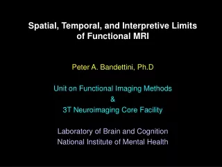
Pushing the Spatial, Temporal and Interpretive Limits of Functional MRI. Peter A. Bandettini, Ph.D Unit on Functional Imaging Methods Laboratory of Brain and Cognition National Institute of Mental Health. Categories of Questions Asked with fMRI. Where? When? How much? ---

E N D
Pushing the Spatial, Temporal and Interpretive Limits of Functional MRI Peter A. Bandettini, Ph.D Unit on Functional Imaging Methods Laboratory of Brain and Cognition National Institute of Mental Health
Categories of Questions Asked with fMRI Where? When? How much? --- How to get the brain to do what we want it to do in the context of an fMRI experiment? (limitations: time, motion, acoustic noise)
A Primary Challenge: ...to make progressively more precise inferences using fMRI without making too many assumptions about non-neuronal physiologic factors.
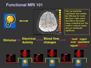
Contrast in Functional MRI • Blood Volume • Contrast agent injection and time series collection of T2* or T2 - weighted images • BOLD • Time series collection of T2* or T2 - weighted images • Perfusion • T1 weighting • Arterial spin labeling
Neuronal Measured Activation fMRI Signal ? ? Hemodynamics Physiologic Factors
Physiologic Factors that Influence BOLD Contrast • Blood oxygenation • Blood volume • Blood pressure • Hematocrit • Vessel size Coupling: Flow & CMRO2
Where and When? The resolution is determined by the cerebral hemodynamics. • Know the vasculature at which you are looking. (or) • Normalize to the spatial variation in the vasculature. (or) • Make several assumptions.
Perfusion / Flow Imaging EPISTAR FAIR . . . - - - - Perfusion Time Series . . .
TI (ms) 200 400 600 800 1000 1200 FAIR EPISTAR
Perfusion Rest Activation BOLD
Anatomy BOLD Perfusion
T1 - weighted T2* weighted T2* and T1* weighted
Spatial Normalization Hypercapnia
, Finger Movement Anatomical 12% O2 5% CO2
Finger Movement Finger Movement / 5% CO2
+ 2 sec Latency - 2 sec Magnitude
Temporal Normalization Relative Timing
Regions of Interest Used for Hemi-Field Experiment Left Hemisphere Right Hemisphere
9.0 seconds 15 seconds 500 msec 500 msec 20 30 10 Time (seconds)
3.2 2.4 1.6 0.8 0 -0.8 -1.6 -2.4 0 10 20 30 Hemi-field with 500 msec asynchrony Average of 6 runs Standard Deviations Shown Percent MR Signal Strength Time (seconds)
3.2 2.4 1.6 0.8 0 -0.8 -1.6 -2.4 0 10 20 30 Average of 6 runs Smoothed Data Percent MR Signal Strength Time (seconds)
500 ms 500 ms RightHemifield Left Hemifield + 2.5 s - = 0 s - 2.5 s
250 ms 250 ms RightHemifield Left Hemifield + 2.5 s - = 0 s - 2.5 s
How Much? Central Issue: Spatial and temporal neuronal firing integration to create an fMRI signal change. - is the hemodynamic response a linear system? -what is the dynamic range?
Motor Cortex Auditory Cortex
2 1000 msec 1.5 100 msec 34 msec 1 0.5 0 -0.5 -1 15 20 25 30 35 Time (sec)
Stimululs - Duration Dependent Deviation from Linearity of the fMRI Response (Hemodynamic or Neuronal?) >linear linear
How to get the brain to do what we want it to do in the context of an fMRI experiment? Noise & Artifact "Interesting" Aspects of MR Signal
Neuronal Activation Input Strategies 1. Block Design 2. Frequency Encoding 3. Phase Encoding 4. Single Event 5. Orthogonal Block Design 6. Free behavior Design.
Overt Word Production 2 3 4 5 6 7 8 9 10 11 12 13
Tongue Movement Jaw Clenching
Event-Related fMRI Questions: • 1. What is the optimal ISI? • 2. How does functional contrast • compare with “blocked” timing? • (Is the hemodynamic response a linear system?)
Contrast in Event Related fMRI • Dependency on: • •Inter-stimulus Interval (ISI) • •Stimulus Duration (SD) • Comparison with: • •Blocked strategies • •Synthesized responses created using • convolution
Issues: • 1. ISI Issue • •Shorter ISI provides more trials per unit time. • •Shorter ISI causes overlap in hemodynamic • response, reducing dynamic range. • 2. Contrast Issue • • Does signal behave like a linear system with • brief SD?
Experimental Methods • •Two imaging planes containing motor and • visual cortex. • •EPI, 3.75 x 3.75 x 7 mm, TE = 40 ms, TR = 1 sec. • •Time series duration = 360 images (6 minutes). • •10 series compared: • Single Trial: SD = 2, ISI = 24, 20, 16, 12, 10, 8, 6, 4, 2. • Blocked: SD = 20, ISI = 20. • •Subjects instructed to tap fingers when GRASS • goggles were on.