Download
1 / 1
10 likes | 101 Views
Calculation of high-resolution and spatially variant photon energy deposition kernels

E N D
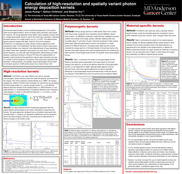
Calculation of high-resolution and spatially variant photon energy deposition kernels Jessie Huang1,2, Nathan Childress3, and Stephen Kry1,2(1) The University of Texas MD Cancer Center, Houston, TX (2) The University of Texas Health Science Center Houston, Graduate School of Biomedical Sciences (3) Mobius Medical Systems, LP, Houston, TX Material-specific kernels Methods: The EDKnrc user code was used to calculate material-specific kernels, except the simulation geometry consisted of various ICRU materials6 (lung, bone, titanium, silver, and gold) rather than water. Results: Table 2 summarizes the results for the material-specific kernels. Notably, the density-weighted effective lateral distance, which indicates how far primary particles travel in the lateral direction (i.e. perpendicular to the direction of the incident photon), is different for different materials and does not simply increase for materials of increasing density. This can also been seen in Figure 3, which shows the angular dose distribution for different material-specific kernels. Polyenergetic kernels Methods: Primary energy spectra of a 6MV photon beam from a Varian 2100 Clinac linear accelerator were calculated using the BEAMnrc Monte Carlo system4. Our BEAMnrc accelerator model has been validated in previous studies5 and consists of the target, primary collimator, flattening filter and moveable upper (Y) and lower (X) jaws. Using this accelerator model, particle phase space data were generated for various scoring planes in a water phantom for different field sizes. This phase space data was then used to calculate the energy spectrum of the beam based on the primary fluence only, and then these beam spectra were used as weighting factors for combining the high-resolution monoenergetic water kernels into spatially variant polyenergetic kernels. Results: Table 1 summarizes the results for the polyenergetic kernels. Based on the beam spectra generated in this study (Figure 2), the mean energy of the spectrum, as well as the effective distances of the polyenergetic kernels, is most dependent on depth. Although depth appears to be the dominant factor, there were spectral differences as well as noticeable differences in the polyenergetic kernels themselves due to field size and off-axis distance. These differences were more pronounced at shallower depths. Introduction Commercial implementations of the convolution/superposition (C/S) method make several approximations, which can lead to dose calculation inaccuracies. For instance, the energy deposition kernel (EDK) used is spatially invariant; that is, a single polyenergetic kernel is used for the entire dose calculation, reflecting the beam spectrum at a single location (e.g. dmax on CAX). This approximation ignores spectral changes with depth, field size, and off-axis distance. Furthermore, for heterogenous media, density scaling is applied to kernels calculated in water. This simplification has been shown to lead to inaccuracies at material interfaces (e.g. water/air)1 and underestimation of dose downstream of metals2.These dose calculation inaccuracies can cause errors in certain clinical situations, e.g. sites with stark heterogeneities (dental fillings and metal implants, lung/tissue interfaces in thoracic RT).Since the implementation of spatially variant kernels has the potential to improve dose calculation accuracy in a variety of clinical situations, the purpose of this study was to generate high-resolution, material-specific, and spatially variant polyenergetic kernels based on the beam spectrum of 6MV photon beam from a Varian Clinac 2100. Figure 3: Normalized dose D(θ)deposited in cones inside the first radial shell (r = 0.05 cm) plotted as a function of the bounding polar angle θfor material-specific kernels simulated with 300 keV incident photons. Expected values of the polar angle <θ> are given in degrees. High-resolution kernels Methods: The EGSnrc user-code2 EDKnrc was used to calculate monoenergetic photon kernels at twice the radial (48 spheres) and three times the angular (144 cones) resolution used by Mackie et al. (1988)3. 22 energies ranging from 100keV to 40 MeV were simulated. For each kernel, we calculated the total energy fraction (Ftot), primary energy fraction (Fprim,), and effective distance along the direction of the incident photon (z), lateral direction (y), and radial direction (r), as illustrated below. The effective distances were calculated using equation (1) where d is the pertinent distance (z, r, or y) and εprim is the primary EDK of the i,j th voxel. (1) Results: Our high-resolution kernels showed good agreement with the original Mackie kernels based on the metrics calculated to characterize the kernels. This good agreement validates that our simulations were performed correctly (Figure 1). However, we did observe differences near the interaction site for the lower energies (<500 keV), most likely due to improvements in electron transport in the EGSnrc code4. Figure 2: Energy spectra of the primary fluence of a Varian Clinac 2100 6MV photon beam for (a) various depths and a 10x10 field, (b) various field sizes at a depth of 1.5 cm, and (c) various off-axis distances for a 20x20 field at a depth of 1.5 cm (using annular scoring planes). Table 2: Density-weighted effective depth of penetration (z), effective radial distance (r), and effective lateral distance (y) for material-specific kernels for 300keV monoenergetic photons and a 6 MeV polyenergetic beam spectrum. In parenthesis is the % difference w.r.t the water kernel. • Conclusions • Our high-resolution water kernels show good agreement with Mackie kernels. However, this good agreement along with the fact that the kernels appear to be smoothly varying functions leads us to believe that they will not appreciably increase the accuracy of dose calculations, with the possible exception of near material interfaces. • For our polyenergetic kernels, we found that depth was the most important factor, but that spectral differences due to field size and off-axis distance were not negligible. • For our material-specific kernels, we found that density scaling is generally a good approximation for lung, but not for higher density, higher effective Z materials such as bone and metals. Use of density scaling for these higher Z materials will lead to underestimation of lateral scatter and dose calculation inaccuracies downstream of such materials. • Based on our data, we expect the use of material-specific kernels and spatially variant polyenergetic kernels to improve dose accuracy for many clinical situations (e.g., downstream of metal implants, in the penumbra region, in peripheral organs at risk, etc). Table 1: Effective depth of penetration (z), effective radial distance (r), and effective lateral distance (y) for polyenergetic kernels calculated using the energy spectrum of the primary photon beam at various locations in a water phantom. The mean energy of the spectrum is also listed. In parenthesis is the % difference w.r.t the polyenergetic kernel at d=1.5 cm for a 10x10 field (i.e. the reference spectrum). Figure 1:Comparison of our high-resolution water kernels and Mackie et al. (1988) kernels averaged over selected angular intervals for (a) 1MeV and (b) 10MeV incident photons. References M. K. Woo and J. R. Cunningham, Med Phys 17 (2), 187-194 (1990). S. Spirydovich, L. Papiez, M. Langer, G. Sandison and V. Thai, RadiotherOncol81 (3), 309-314 (2006). D. W. O. Rogers, I. Kawrakow, J. P. Seuntjens, B. R. B. Walters, and E. Mainegra-Hing, Technical Report PIRS-702(RevB), National Research Council of Canada, Ottowa, Canada (2003). I. Kawrakow, E. Mainegra-Hing, D.W.O Rogers, F. Tessier, and B. R. B. Walters. Technical Report PIRS-701. National Research Council of Canada, Ottowa, Canada (2011). T. R. Mackie, A. F. Bielajew, D. W. Rogers and J. J. Battista, Phys Med Biol33 (1), 1-20 (1988). D. W. O. Rogers, B. Walters, and I. Kawrakow, NRCC Report PIRS-0509(A)revK, National Research Council of Canada, Ottawa, Canada (2009). O. N. Vassiliev, U. Titt, S. F. Kry, F. Ponisch, M. T. Gillin and R. Mohan, Med Phys 33 (4), 820-827 (2006). ICRU, ICRU Report 37, ICRU, Washington D.C. (1984). Support The investigation was supported by PHS grantCA10953 awarded by the NCI, DHHS. Contact: jyhuang@mdanderson.org