Download
1 / 71
740 likes | 784 Views
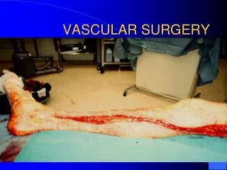
VASCULAR SURGERY. Raynaud's. Raynaud’s phenomenon the phenomenon is the spasm in response to cold or emotion (pallor, cyanosis ruber sequence is not essential for diagnosis) Raynaud’s syndrome, is the phenomenon associated with another disease

E N D
Raynaud's • Raynaud’s phenomenon the phenomenon is the spasm in response to cold or emotion (pallor, cyanosis ruber sequence is not essential for diagnosis) • Raynaud’s syndrome, is the phenomenon associated with another disease • Raynaud’s disease is phenomenon occurring primarily in isolation
Pathophysiology Neurogenic • Oversensitivity of alpha adr rec. • Query involvement of central sympathetic system Blood borne factors • Blood and vessel wall interaction • RBC, WBC wall rigidity-->aggregation • Vasoactive hormones secretion is disturbed (endothelin, PGI2, TXA2) Inflammatory and immunological mechanism The firm association with CTD (connective tissue diseases)
Clinical features Diagnosis can usually be made from clinical history, Consists of a triphasic response provoked by exposure to cold Phase 1 - pallor due to intense vasoconstriction Phase 2 - cyanosis due to desaturation of haemoglobin Phase 3 - erythema due to hyperaemia and restoration of circulation • Primary disease is usually bilateral, symmetrical and involves all fingers • Secondary disease is usually patchy and asymmetrical • Symptoms are often milder in primary disease • Examination often shows peripheral pulses to be normal • Features of thoracic outlet syndrome or connective tissue disorders may be present
DD • Ergotism • Acrocynosis • Livedoreticularis • Scalene syndrome
Investigations • Should be guided by clinical features • No reliable method of provoking Raynaud's phenomenon • Start with laboratory tests • Full blood count, urea, electrolytes, and urine analysis ------->chronic renal disease • Thyroid function test to exclude hypothyroidism • ESR, plasma viscosity, rheumatoid antibody and antinuclear antibody------->CTD • Duplex ultrasound or arteriography may be indicated if suspicion of arterial disease exists –Confirm diagnosis (diagnosis is usually only clinical) –Differentiate between primary and secondary –Elucidate the underlying disease
Conditions associated with Raynaud’s phenomenon • Connective tissue diseases • Systemic sclerosis • Systemic lupus erythermatosus • Rheumatoid arthritis • Sjogren syndrome • Obstructive • Atherosclerosis • Microemboli • Thoracic outlet syndrome specially cervical rib • Drug therapy • B blockers • Cytotoxic , specially bleomycin • Cyclosporin • Antimigrane therapy
Conditions associated with Raynaud’s phenomenon • Occupational • Vibration white finger syndrome • Frozen food packers • Ammunition workers • Miscellaneous • Hypothyroidism • Cryoglobinemeia • Malignancies
hyperhydrosis pallor
Treatment options Prevention • Clothing - wearing of thermal fabrics • The use of hand warmers or electric gloves • Stop smoking • Change job if vibration induces • Avoid sympathetic stimulants Topical agents • GTN paste • Prostaglandin analogues
Treatment options Oral agents • Sympatholytic agents -reserpine, guanethidine • Alpha-blockers - phenoxybenzamine, prazocin • Beta-stimulants - terbutaline • Vasodilators - nifedipine, diltiazen • Fibrinolytic agents - pentoxifylline • Serotonin antagonists - ketanserin Invasive • Intraarterial reserpine • Intravenous iloprost
Treatment options Surgery • Thoracic outlet surgery • Embolectomy • Sympathectomy • Lumbar sympathectomy Important role in intractable RP of the feet and is often worthwhile trying • Cervical sympathectomy •Has a poor response, and high relapse •It is no longer used for upper limb RP
Buerger’s Disease • It is episodic, segmental , Inflammatory and thrombotic process • affecting arteries and veins • mainly in legs in male , around 40 years and smoker
Buerger’s Disease ( etiology) • Immunological • Collagenic • Sympathetic over activity • Disorder of blood clotting
Buerger’s Disease ( pathology) • Arteries • Small and medium size • LL > UL • Veins • Thrombophelibits • UL> LL • Nerves • Raynaud’s phenomenon
Buerger’s Disease ( Clinical) • Mostly in male, 40, smoker • Progressive episodic course • Take 4 stages • Phelibitis migrans • Intermittant claudication • Rest pain • Trophic changes
Buerger’s Disease ( Investigation) • Plain x ray no calcification • Arteriography • Small and medium sized • Smooth intimal lining • Cork scrow collaterals, foot tree or spider like • Echo to exclude cardiac emboli
Treatment • Non surgical • Prevent smoking • Vasodilators • Corticosteroids in attacks • Surgical • Sympathectomy • Amputations
Arteriovenous fistulsa Communication between an artery and a vein may be either • Physiological AVF • Congenital • Traumatic • Therapeutic • Pathological • BHF, liver cirrhosis • Angidyspasia • Paget’s disease of bone
Arteriovenous fistulsa Congenital malformation(high flow vascular malformation) these are clinically divided into three groups based on structural viz • Truncal (major arterial branches mainly upper limb and head and neck) , • Diffuse (more in the lower limb) , and • Localized (in hands or feet or organs).
Arteriovenous fistulsa Clinically • Distally • Veins 2 ry VV • Arteries Ischemia • Local • These are characterized by warmth ,palpable thrill and an audible bruit. • Growth of the limb gigantism • Central cardiac enlargement Angiography or MRA clinch the diagnosis . Treatment • Non surgical embolisation, sclerosent agent, cogulation transcatheter • surgical excision are the lines of treatment.
Arteriovenous fistulsa Acquired by • Trauma of a penetrating wound • Created surgically in patients undergoing dialysis .It is known as Cimino’s shunt. • Fracture skull base • Iatrognic
Arteriovenous fistulsa Clinically • Distally • Veins 2 ry VV • NO Arteries Ischemia • Local • These are characterized by warmth ,palpable thrill and an audible bruit ( systo-diastolic). • NO Growth of the limb gigantism • Central • Tachycadia • Bradycardia on compression ( Branham’s sign) • Water hammer pulse • Heart failure and hypertrophy
Arteriovenous fistulsa Aortocaval fistula
Treatment For congenital types • embolisation by the radiologist, or • excision is advocated only in severe deformity or recurrent hemorrhage • Ligation of the feeding artery is of no lasting value, but can impair treatment with embolisation The acquired lesions are best treated when small because they are progressive • sometimes embolisation can cure the case • In operation after control of the artery proximal and distally • the communication can be repaired surgically. • Four ligatures for the artery and vein involved (quadrable ligation ) can be used.. • Also bypass graft can be the solution.
Diabetic • Diabetic Neuropathy • Sensory • Moor • Autonomic • Vascular abnormalites • Infections • CT abnormalites • Hematolgical abnormalites
Neuropathic pain Intermittent claudication Ischemic rest pain Foot/shin Foot/calf Calf/thigh Tingling/burning/shooting Aching Cramping Increase by Night time Elevation Exercise Decrease by Exercise Dependency of foot Rest Warm, bounding pulses Weak, absent pulses Cold/pulseless 1Comparison of signs and symptoms of neuropathic and ischemic pain