Download
1 / 41
420 likes | 572 Views
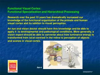
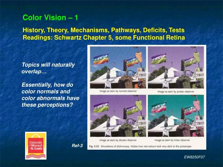
Color Vision – 1 History, Theory, Mechanisms, Pathways, Deficits, Tests Readings: Schwartz Chapter 5, some Functional Retina. Topics will naturally overlap… Essentially, how do color normals and color abnormals have these perceptions?. Ref-3. EW8250F07.

E N D
Color Vision – 1History, Theory, Mechanisms, Pathways, Deficits, TestsReadings: Schwartz Chapter 5, some Functional Retina Topics will naturally overlap… Essentially, how do color normals and color abnormals have these perceptions? Ref-3 EW8250F07
Another simulation of color normal and color abnormal perceptions. Ref-4 EW8250F07
Neurophysiological Example of Color ConstancyArea V4 neurons show this property whereas V1 neurons do not. Green tuned V1 and V4 neurons. Only the V1 neuron fires when green illuminates the white square. Thus color constancy is a property of only the V4 neuron. Ambient green is subtracted only in V4 Ref-3 EW8250F07
Example of Simultaneous Color ContrastThe grays are physically equal but take on the hue of the surround. Ref-4 EW8250F07
Examples of Successive Color ContrastColor adaptation effect from extended viewing of the left image. Ref-3 Ref-4 EW8250F07
Wavelengths / Colors(not all figures agree)420 nm Violet470 nm Blue500 nm Blue-Green520 nm Green555 nm Yellow-Green570 nm Yellow600 nm Orange650 nm Red507 nm Peak scotopic (rod) sensitivity555 nm Peak photopic (cone) sensitivity Ref-4 Ref-3 EW8250F07
Source and Surface Factors Influence Color Perception Same apple but different color appearance per Illuminant. Appears more red with illuminant A. EW8250F07
Hering incorrectly hypothesized that opponent channels arose from each cone having two photopigments in opposition: one built up (anabolized) and one destroyed (catabolized) in any light. EW8250F07
Basic Scheme: Red/Green, Blue/Yellow and Luminosity Outputs Ref-3 EW8250F07
Opponent Processing (R/G, B/Y, B/W)It may help by studying, and you should be aware of,the associated pathways from functional retina. For example: Ref-5 Midgets for R/G Parasols for R/G and B/W S-cone cells link to M- and L-cones (labeled ‘LM’) for B/Y EW8250F07
Production of ColorThere are three ways to produce color:Colored LightsSubtractive MethodAdditive MethodProduction of Color: Colored lightsElectric discharge lamps emit light of a single wavelength or a few wavelengths, e.g. sodium discharge lamp and mercury vapor lamp, both used for street lighting.Lasers emit a single wavelength. EW8250F07
Example of Subtractive Method Using PigmentsA mixture of blue and yellow pigments appears green because blue absorbs long wavelengths and yellow absorbs short wavelengths. What reaches the eye are only middle wavelengths, the only band reflected by both pigments. Ref-8 This pigment condition is analogous to passing light through a blue and a yellow filter. EW8250F07
Additive MethodLight(s) shown,cones activated,and resultantcolor perception. Ref-8 EW8250F07
Additive and Subtractive MethodsAdditive primaries: blue, green and red.Subtractive primaries: cyan, magenta and yellow. Ref-3 EW8250F07
Examples of Additive and Subtractive Methods Using FiltersAdditive: B + Y = WSubtractive: B + Y = G Ref-4 EW8250F07
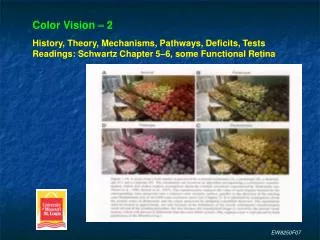
Color Vision – 2History, Theory, Mechanisms, Pathways, Deficits, TestsReadings: Schwartz Chapter 5–6, some Functional Retina Ref-3 EW8250F07
1.2 1.0 0.8 Relative sensitivity 0.6 0.4 0.2 0.0 400 425 450 475 500 525 550 575 600 625 650 675 700 Wavelength (nm) The 3 general photopigment, or cone, classes have broadly overlapping ranges of spectral sensitivity (absorption). Cut-offs to note: ~ 525 nm ~ 650 nm EW8250F07
Homology (identical DNA) exists between the gene of rhodopsin and those of the cone photopigments. This suggests a common ancestor of all 4 photopigments. The strongest homology is between rhodopsin and S-cones.M- and L-cone homology (identical DNA) = 98% = extremely strong.Suggests that both evolved recently.M- and L-cone to S-cone homology = 40% = not strong.Suggests that the S-cone evolved earlier. Pink = amino acids unequal EW8250F07
Ripe fruit in green background:evolutionary argument forthe development (i.e. need)of the L-cone photopigment. Area V8 is another higher-order color center. EW8250F07
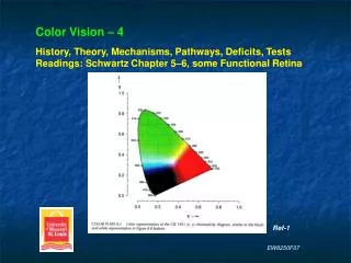
Color Vision – 4History, Theory, Mechanisms, Pathways, Deficits, TestsReadings: Schwartz Chapter 5–6, some Functional Retina Ref-1 EW8250F07
Tilted lines are hue contour lines.They show the wavelengths that keeps a constant hue (to the reference) with increasing luminance.The 3 invariant wavelengths are478 nm, 503 nm and 578 nm.These are also called invariantpoints or unique hues. They lookpure in that they do not appear tobe mixed with another wavelength. 478 nm = unique blue 503 nm = unique green 578 nm = unique yellow Ref-2 EW8250F07
Munsell Color Appearance SystemThese colors are presented in a Book of Color as chips.The color of any surface can be identified by comparison to the chips then assigned a H V/C value.The 3-dimensions are often represented as a color solid. EW8250F07
Design of the Munsell Color Appearance System.The three attributes (dimensions) of color are perceptual attributes. Hue = perimeter, 10 wavelength bands (RYGBV), 0 – 100 Value = top to bottom in center, brightness 0 – 10 Chroma = radius, saturation, 0 – 14 (some 0 – 30) EW8250F07
Example H V/C for vivid red: 5R 6.2 / 13.5 (wavelength, brightness, saturation The Munsell Color Appearance System is widely used in industry, i.e. the public. EW8250F07
CIE Color Specification SystemLike the Munsell System, it is designed to specify colors. However, this system is used in vision research and for clinical applications.Based on trichromatic vision: any color is specified by the relative amounts of a mixture of three primary colors.It is a system of imaginary primaries (X, Y and Z) developed because of the difficulty in using red, green and blue primaries. Ref-1 EW8250F07
Note how combinations of chromaticity coordinates produces specific colors. EW8250F07 Ref-2
CIE Diagram Color Confusion Lines Similar Different Ref-2 Ref-1 EW8250F07
CIE Diagram and the D15 and 100-hue Cap Tests 15 cap test 85 cap test Ref-1 Ref-2 EW8250F07
ColorLabeling Ref-2 EW8250F07
Color Labeling Ref-2 EW8250F07
Color Vision – 5History, Theory, Mechanisms, Pathways, Deficits, TestsReadings: Schwartz Chapter 5–6, some Functional Retina • Nice depiction of better • color discrimination with • additional photopigment. • Fruit photo • Monochromat • Dichromat • Trichromat • Would a tetrachromat • (contribution from rods • or, say, female carriers) • perceive colors better? • More to the point, does • tetrachromatism come • with added (i.e. required) • post-receptor opponent • processing? EW8250F07
Ganglion Cells: vast evidence for color opponency; although much is unknown about how the surround (M+L versus onlyM or L) forms in the R/G channel. Where are color opponent cells first found? Retina and LGN. Where are more color opponent cells found? Cortex > LGN > Retina. Where are double color opponent cells first found? V1 and V2. EW8250F07
Older schematic using B, R and G rather than S, M and L. R+/B– Does Not Exist EW8250F07
In view of the principle of univarianceand the limits of retinal mechanismsto date, it seems likely that centralmechanisms (LGN and visual cortex)elaborate the wavelength-specific(chromatic) information embeddedin cone signals.Natural scene to retinal image.S-, M- and L-cone quanta absorptions.No λ information transmitted by cones(Principle of Univariance).Opponent processing and their specific combinations in the retina and LGN.3 channels (B/W, R/G, B/Y) in retina andopponent ganglion cells and LGN cells.Not shown: further / finer processing inearly and higher visual cortex. Trichromacy Cone output: Opponency Opponent output: EW8250F07
With regard to transmitting chromatic and achromatic information…Magnocellular (M) cells: transmits achromatic, or luminance, informationParvocellular (P) cells: transmits R/G color opponent informationKoniocellular (K) cells: transmits B/Y color opponent informationLGN: M-cells synapse in layers 1 and 2, P-cells in layers 3 – 6, andK-cells in the interlaminar (6) spaces. 6 K-cell layers, one below each M- and P-layer. EW8250F07
Neurophysiological Example of Color ConstancyArea V4 neurons show this property whereas V1 neurons do not. Green tuned V1 and V4 neurons. Only the V1 neuron fires when green illuminates the white square. Thus color constancy is a property of only the V4 neuron. Ambient green is subtracted only in V4 Ref-3 EW8250F07
Area V8Deficit gives cerebral achromatopsia.Many documented stroke cases.End-stage ‘color perception’ area?Strong evidence from after-image studies: neural activity is seen during the after-image, i.e. when seeing the illusion of color.Acuity and most other visual abilitiesare maintained – no retrograde affects. Ref-3 EW8250F07
Area V8 is located very close (just anterior) to area V4.A ‘V4/V8 color complex’ is becoming commonly referred to in research. Ref-3 Area V8 Area V4 Ref-3 EW8250F07
Add Slide:Mechanism for the perception of white (neutral point) in dichromats. white wavelengths Ref-4 EW8250F07