Download
1 / 20
200 likes | 232 Views
Iliotibial band (ITB) syndrome is one of the most common overuse injuries in runners. Virtual physical therapy helps runners identify the biomechanical issues of ITB and educates them with knowledge about the causes of iliotibial band syndrome, biomechanics, causes of pain, misdiagnosis, rehabilitation, prevention, and more.

E N D
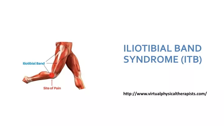
ILIOTIBIAL BAND SYNDROME (ITB) http://www.virtualphysicaltherapists.com/
Iliotibial band (ITB) syndrome is one of the most common overuse injuries in runners. It is also seen in cycling and other activities that require repetitive bending of the knee. Treatment must focus on eliminating the causative factors. Virtual Physical Therapy can help identify biomechanical issues and educate you on how you can get back to running pain-free!
ITB syndrome usually starts suddenly as discomfort or even burning on the outside of the knee. It can quickly turn into a sharp pain and quickly progress to a feeling of a vice around your knee that gets tighter and tighter until the pain stops you from moving. Initially, the symptoms are only brought on by aggressive activity such as running and cycling but as they progress, sitting with your knee flexed becomes unbearable, requiring straightening of the knee to release pressure.
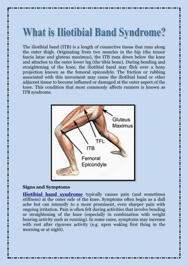
What Causes Iliotibial Band Syndrome? The number one cause is too many miles! Minor issues with running mechanics become exacerbated when fatigued, and your muscles are weakened from overtraining. Not allowing your body time to heal leads to poor mechanics, tears in soft tissue, damage. In the case of ITB syndrome it is due to irritation and thickening of a fat pad over the femoral epicondyle that the ITB repetitively compresses. Banked surfaces (always running in the same direction as road camber) and downhill running (eccentric muscle control gives way faster when fatigued) can cause increased stress and compression of the ITB.
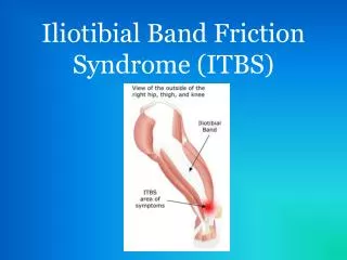
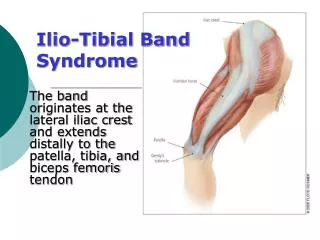
Anatomy: The ITB is a thick fibrous band extending from above your hip to below your lateral knee. The proximal portion begins as a sheath encasing the tensor fascia lata muscle. This sheath anchors the tensor fascia lata to the iliac crest and receives most of the superior gluteus maximus tendon. The dense ITB then extends all the way down the lateral leg and transitions to a ligamentous component, spanning from the lateral epicondyle of the femur (just above the outside knee) to five insertion points distally including the knee capsule and Gerdy’s tubercle just below the lateral knee.
Biomechanics: The ITB acts as both a stabilizer for the hip and knee as well as elastic energy during walking and running. It is a distinctive tissue in that it has some properties of a tendon but most of a ligament. It is also unique because humans are the only mammal to have one. It is theorized that the ITB aids in our ability to stand on one leg and walk upright and is not needed in 4 legged mammals. We are not born with a distally inserting ITB, but rather developed as we learn to walk. The iliotibial band transmits the forces generated by the TFL and gluteus maximus muscles, including thigh abduction, flexion, extension, and external rotation. The deep fascial component, which runs almost the entire femur length, is most taut when the gluteus maximus and TFL contract. This “tensile” action significantly increases during single-leg stance and serves to counteract medial bowing of the femur.
The ITB is actually a ligament/tendon hybrid because it has a bone-to-bone connection as seen in ligaments and muscle-to-bone connections as found in tendons. The ITB is NOT anchored to a bone at a clear, specific spot like most tendons. Instead, it blends into five different areas distally including the knee capsule. Tendons are smaller, dense connective tissue at the end of the muscle. The ITB, on the other hand, connects the muscles by a very thin sheath and is massive compared to the muscles it connects. The gluteus muscle pulls on the ITB to increase its tension laterally, like drawing a bowstring. It is then tightly anchored along the entire length of the femur, especially above the knee. The ITB does not really move freely in relation to the femur. It is firmly attached. The only slight movement may be at the insertion at Gerdy’s tubercle, just below the lateral knee.
Cause of Pain: Iliotibial band syndrome occurs secondary to repetitive compression. Faulty mechanics caused an increase in compression at the distal end of the ITB as the hip extends and the knee flexes. The bony prominence of the lateral epicondyle has a protective synovial tissue or fat pad. This synovial tissue can become irritated, thickened, and even calloused. Once calloused, it can be felt like rubbing over a rubber band.
For many years it was believed that the pain was due to ‘friction’ of the ITB over the lateral femoral epicondyle. The ITB is tethered to the distal femur, except for the upper portion of the lateral femoral condyle, preventing movement of the ITB across the lateral femoral condyle. Biomechanics that cause an increase in the compression of the ITB include genu varum (knees go inward), increased pronation, and hip weakness. These are in opposition to the role of the ITB, to pull on the femur laterally like a bowstring. The main problem and symptoms occur at 30 deg of knee flexion with the hip in slight extension – this is when the ITB is mostly clamped down.
Misconceptions: 1. The ITB is the painful structure. The painful tissue is underneath the ITB. It is synovium or tissue similar to a fat pad that is a lateral extension of the knee capsule. Repetitive stress causes it to become callused, and it gets “pinched”. Occasionally there can also be calcified loose bodies if the stress on this synovial tissue continues. 2. The ITB needs to be stretched using a foam roller or specific stretching. The ITB is mostly a ligament in structure. The role of a ligament is to maintain a taunt attachment to bone. You do not want loose ligaments as this leads to instability. The ITB is also a vast thick structure similar to a thick leather belt. You can pull on it all you want, and it still will not stretch. Foam rolling adds more compression to a tissue that has been already irritated by compression.
3. Corticosteroid injections – causes local cell death and tendon atrophy! Steroids have an important place in medicine but must be used with caution. Inflammation is part of the body’s natural healing process, and when it is terminated, it disrupts healing and can lead to tendon damage. The main treatment focus should be addressing the actual cause of ITB syndrome or repetitive stress/compression causing the tissue to become calloused.
Running: Humans are built for running, and we may even be one of the best species for long-distance. Every year in Wales, there is an endurance race of humans against horses, and believe it or not, humans won twice because of hot conditions. If we are built for running, then why do we get so many injuries? It’s because of poor mechanics and our training.
Running: Humans are built for running, and we may even be one of the best species for long-distance. Every year in Wales, there is an endurance race of humans against horses, and believe it or not, humans won twice because of hot conditions. If we are built for running, then why do we get so many injuries? It’s because of poor mechanics and our training.
Mechanism of Injury: The ITB is most taunt when the hip is extended and the knee is flexed to 30 degrees. The hip goes into extension during the swing phase of running when the knee bends. Severe symptoms almost completely abolish as soon as the individual stops walking because the hip does not extend in walking. The faulty mechanics found in those that suffer from ITB on the painful side include 1. Hip drops down and in (adducts) 2. Ankle bone drops down and in (pronation) during heel strike 3. Heel is inward during the swing phase.
The faulty mechanics are exacerbated with fatigue as the muscles tire leading to weakness in hip abduction weakness causing the hip to drop inward and tibialis anterior fatigue causing pronation of the foot. Road cadence – constantly running on one side of the road places uneven stress on the leg, and poor footwear can cause increased pronation.
Physical Evaluation: 1. A good clinician will first RULE OUT the lumbar spine. There is a high incidence of isolated extremity symptoms originating from the spine. Therefore the spine must always be screened. This is easily done by moving the spine to see if it has any effect on the symptoms of complaint along the outer knee. 2. Observation: Screen for alignment and any abnormalities 3. Palpation: Assess for any tenderness and palpable bursa along the lateral femoral eopicondyle. 4. Test for ITB Syndrome: Have the individual repetitively flex and extend the knee with hip in extension. A positive test is the reproduction of lateral knee pain. 5. Muscle strength (hip abduction, external rotation, quad, foot) 6. Flexibility: quad, hamstring, calf, and soleus 7. Gait/running assessment
Rehabilitation: • Active REST • An initial rest from running in favor of another aerobic activity – cycling, swimming, etc. so activity can continue while eliminating compression over the lateral epicondyle. Gradually return to running (initially avoid downhill) 3 days to 6 weeks depending on the individual’s symptoms. Average avoidance of running – 1 week. (2-6 months until recovery is complete) • Address limitations • Full quad, hamstring, and calf flexibility • Improve hip, knee, and foot strength
Address running mechanics • Land with ankle bone high • Swing phase with heel out • Return to running • Faster, shorter runs initially (Slow running causes increased pressure on ITB. Sprinting reduces compression.) • Adjust running style – shorter stride and lower (initially) • Avoid downhill • Gradually increase millage
Prevention: • Learn proper running mechanics (GOATA). Schedule an assessment with one of our GOATA specialists. • Training!!!! Gradual increase in millage • Maintain adequate strength and flexibility • Avoid always running on one side of the road if there is camber
Virtual physical therapists info.virtualphysicaltherapists@gmail.com http://www.virtualphysicaltherapists.com/