Download
1 / 27
350 likes | 618 Views
Mouse as a Model Organism. Tuesday, February 7, 2012. Overview. Reproduction Grafting Non-Homologous Recombination Homologous Recombination Cre / loxP Recombination Tissue Growth Histology Models of Human Disease. Reproduction. 5-10 litters / year 5-10 pups / litter

E N D
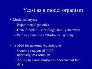
Mouse as a Model Organism Tuesday, February 7, 2012
Overview • Reproduction • Grafting • Non-Homologous Recombination • Homologous Recombination • Cre / loxP Recombination • Tissue Growth • Histology • Models of Human Disease
Reproduction • 5-10 litters / year • 5-10 pups / litter • 19-21 day gestation • Sexually mature at 7 weeks • 4-5 generations per year
Grafting • Cannot do it! • Cells are too small
3 Types of Genetic Modifications • Insertion – of a transgene or a modified allele, i.e., “knock-in” – can produce a gain of function mutation • Knockout – of a particular gene or piece of DNA – to assess a gene’s function, i.e., is it necessary for a particular role in development • Conditional Mutant – a spatially and temporally specific knockout!
I. Inserting DNA into Cells • 1) Microinjection of cloned gene into nucleus of newly fertilized egg • 2) Transfection incubate ES cells in solution that makes them take up the DNA, very inefficient need to identify cells that took up the DNA with reporter such as drug resistance • 3) Electroporation – a high voltage pulse “pushes” DNA into cells • 4) Retroviral vectors – a more natural way or getting genes into cells
Electroporation Highly efficient for the introduction of genes in mammalian tissue culture cells
II. Knocking out a gene • Homologous recombination • Clone gene that is nonfunctional • Introduce DNA into cell by any method discussed above • Homologous recombination will occur replacing endogenous gene with nonfunctional gene
Conditional Mutant: Cre-LoxP • Conditional mutants are needed when you want to study the effects of a gene in certain tissue late in development but the gene is also necessary early in development. A traditional knockout would result in a mutant that does not develop to stage needed. • Cre is a recombinase that excises DNA located in between LoxP sites • You generate two transgenic lines one that expresses Cre in the tissue you are interested and a second that contains gene of interest flanked by loxP sites. The gene will only be deleted where Cre is expressed. • Can also activate genes: In second line place stop signal flanked by loxP between 5’ regulatory element and gene. When stop signal is removed gene will be activated.
Tissue Culture • Possible to grow certain tissues in vitro • Need to have isolated stem cell line • Most tissue type has different protocols • Does not form functioning organ
Embryo and Organ culture • Can remove entire embryo or organ and maintain alive in culture for a short period of time • Add factors to embryo or organ: activators, inhibitors, drugs • Afterwards do whole mount or sections in situ hybridization, RT-PCR, immunostaining ect. to analyze the embryo or organ. • Can also do tissue transplantations • Can also remove at different stages to observe development
Histology • Immunohistochemistry (Antibody staining) • In situ hybridization • Cell death staining • Bone and cartilage chemical stains
Cell Death Staining • TUNEL (Terminal deoxynucleotidyl transferase dUTP nick end labeling), Nile Blue, Acridine Orange • Used to detect apoptosis • Tunel: detects DNA fragmentation by labeling the ends of the DNA TUNEL Nile Blue Acridine Orange
How to detect Cell Proliferation • BrdU is a synthetic nucleoside that is an analogue of thymidine. BrdU is commonly used in the detection of proliferating cells in living tissues. • BrdU labeling: (Bromodeoxyurdine) BrdU incorporated into cells that are undergoing DNA synthesis. Detected with antibody staining.
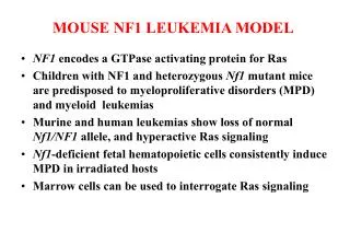
Model of Human Disease • Many known gene mutations exist that reproduce human diseases in mice. • Are these accurate models of human disease? • Not all mouse phenotypes correspond to human phenotypes • Studies primarily done in C57BL/6 strain • Is a study of a single strain sufficient to make conclusions about humans?