Download

1 / 36
360 likes | 389 Views
Question & Answer session - Quiz with keepads.

E N D
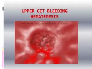
Pathology of Upper GIT - Quiz He who is not courageous enoughto take risks will accomplish nothingin life.– Muhammad Ali, Champion Boxer
A. B. C. D. E.17%8%67%0%8%? diagnosisA. Adenocarcinoma.B. Acute Oesophagitis.C. Barrett’s oesophagusD. Oesophageal varicesE. Achalasia A. B. C. D. E.17%8%67%0%8%? diagnosisA. Adenocarcinoma.B. Acute Oesophagitis.C. Barrett’s oesophagusD. Oesophageal varicesE. Achalasia
A. B. C. D. E.33%0%11%56%0%42 year old man, chronic alcoholic, develops intractablehematemesis and ultimately exsanguination. Au autopsy, theopened up oesophagus appeared like the image. What is themost likely cause?.A. Forced vomiting.B. Columnar metaplasia.C. Malignant change.D. Portal hypertension.E. Reflux of gastric acid. A. B. C. D. E.33%0%11%56%0%42 year old man, chronic alcoholic, develops intractablehematemesis and ultimately exsanguination. Au autopsy, theopened up oesophagus appeared like the image. What is themost likely cause?.A. Forced vomiting.B. Columnar metaplasia.C. Malignant change.D. Portal hypertension.E. Reflux of gastric acid.
A. B. C. D. E.0%69%13%0%19%65year old man with long standing GORD now has a 2month history of hematemesis and dysphagia. His loweroesophagus gross & Microscopy appears similar to theimage. What is the most likely complication?.A. Acute oesophagitis.B. Barrett’s oesophagus.C. adenocarcinoma.D. Squamous cell carcinoma.E. Perforation. A. B. C. D. E.0%69%13%0%19%65year old man with long standing GORD now has a 2month history of hematemesis and dysphagia. His loweroesophagus gross & Microscopy appears similar to theimage. What is the most likely complication?.A. Acute oesophagitis.B. Barrett’s oesophagus.C. adenocarcinoma.D. Squamous cell carcinoma.E. Perforation.
46y male chest pain. Oesophageal biopsy.A. B. C. D. E.7%50%21%0%21%A. Chronic esophagitisB. Squamous metaplasiaC. Barrett’sD. Adenocarcinoma.E. Squamous carcinoma 46y male chest pain. Oesophageal biopsy.A. B. C. D. E.7%50%21%0%21%A. Chronic esophagitisB. Squamous metaplasiaC. Barrett’sD. Adenocarcinoma.E. Squamous carcinoma
58y Fem, hematemesis and hematochezia, alcoholiccirrhosis 2y ago, Lower oesophagus biopsy. What is themost likely Diagnosis?1 2 3 4 531%13%6%0%50%1. Mallory Weiss Syndrome.2. Barrett’s esophagus.3. Esophageal varices4. Sliding Hiatus hernia5. Acute esophagitis. 58y Fem, hematemesis and hematochezia, alcoholiccirrhosis 2y ago, Lower oesophagus biopsy. What is themost likely Diagnosis?1 2 3 4 531%13%6%0%50%1. Mallory Weiss Syndrome.2. Barrett’s esophagus.3. Esophageal varices4. Sliding Hiatus hernia5. Acute esophagitis.
56y male, abdominal pain, Gastric biopsy, ? arrowA. B. C. D. E.0% 0%6%94%0%A. Barrett’s.B. H.pylori gastritis.C. Chronic gastritisD. AdenocarcinomaE. Gastric metaplasia. 56y male, abdominal pain, Gastric biopsy, ? arrowA. B. C. D. E.0% 0%6%94%0%A. Barrett’s.B. H.pylori gastritis.C. Chronic gastritisD. AdenocarcinomaE. Gastric metaplasia.
46y male pain, hematemesis: Stomach.1 2 3 4 511%89%0%0%0%A. Malignant Gastric ulcerB. Benign peptic ulcer.C. Barrett’sD. Adenocarcinoma.E. Acute gastritis. 46y male pain, hematemesis: Stomach.1 2 3 4 511%89%0%0%0%A. Malignant Gastric ulcerB. Benign peptic ulcer.C. Barrett’sD. Adenocarcinoma.E. Acute gastritis.
46y male pain, hematemesis: Stomach.1 2 3 4 50% 0% 0%0%0%A. Malignant Gastric ulcerB. Benign peptic ulcer.C. Barrett’sD. H.pylori gastritis.E. Gastric Perforation. 46y male pain, hematemesis: Stomach.1 2 3 4 50% 0% 0%0%0%A. Malignant Gastric ulcerB. Benign peptic ulcer.C. Barrett’sD. H.pylori gastritis.E. Gastric Perforation.
. 46y male odynophagia : Esophageal biopsy.A. B. C. D. E.0% 0%13%13%73%A. Barrett’sB. Acute Esophagitis.C. Squamous CarcinomaD. Adeno Carcinoma.E. Chronic Esophagitis.
. Correct statement about H.pylori?1 2 3 4 523%62%8%8%0%A. Gram positive spirocheteB. Colonizes Gastric mucosaC. Invades duodenal mucosaD. Diagnosed by bacterial culture.E. Complication is Duodenal cancer.
. Common site of Peptic Ulcer?1 2 3 4 518%0%73%9%0%A. Cardiac part of Stomach.B. Greater curvatureC. Lesser curvatureD. 2nd part of duodenumE. 1st part of duodenum
. 46y male odynophagia : Esophageal biopsy.A. B. C. D. E.0% 0% 0%0%0%A. Barrett’sB. Acute Esophagitis.C. Squamous CarcinomaD. Adeno Carcinoma.E. Chronic Esophagitis.
. 35y man, chronic dysphagia, regurgitatefood. Endoscopy normal. Flow studies showlack of peristalsis. ? Diagnosis?.1 2 3 4 512%53%18%18%0%1. Schatzki ring.2. Achalasia3. Barrett oesophagus4. Esophageal stricture.5. Mallory-Weiss sy.
. 46y male chestpain,Endoscopy:oesophagus1 2 3 4 50%17%25%8%50%A. Hiatus herniaB. Acute Oesophagitis.C. Barrett’s oesophagusD. Oesophageal varicesE. Mallory weiss sy.
. 20y fem, 2y history of dysphagia, fatigueand pallor, microcytic RBC ? Diagnosis?.1 2 3 4 531%15%8%23%23%1. Barrett esophagus2. Diverticulum3. Esophageal web4. Schatzki ring5. Achalasia
. 76y man, Sudden-onset deep burning epigastric painradiating to abdomen for 4 hours. Past history of coronaryartery disease, hypertension & “indigestion.” Hyperactivebowel sounds are heard on auscultation. Xray abdomen,? diagnosis1 2 3 4 517%0% 0%0%83%1.Aortic aneurysm rupture2.Acute Pancreatitis3.Pneumoperitoneum PUD4.Acute coronary syndrome5.Ruptured MI
. 46y male odynophagia : Esophageal biopsy.A. B. C. D. E.0% 0% 0%83%17%A. Barrett’sB. Acute Esophagitis.C. Squamous CarcinomaD. Adeno Carcinoma.E. Chronic Esophagitis.
. 34y Male, Insomnia, heart burn, dysphagia.Lower Esophagusendoscopy:• Features ?• Etiology ?• Clinical features ?• Complications ?
. 45y female, attacks of wheezing, SOB, hot flashes. Abdominalcramps, diarrhoea, Tricuspid regurgitation, Increased urinary 5-HIAA, CT scan shows nodule in jejunum, stomach and fewnodules in liver. ? Diagnosis.1 2 3 4 58%46%15%23%8%1. GIT Lymphoma.2. Carcinoid tumor.3. Mallory Weiss Syndrome.4. Gastric carcinoma with mets.5. Zollinger Ellison syndrome.
. “Only a man who knows what it is liketo be defeated can reach down to thebottom of his soul and come up with theextra ounce of power it takes to win,when the match is even.”– Muhammad Ali, Champion Boxer
. 56y male, hematemesis: Gastric biopsy, ? arrowA. Barrett’s.B. H.pylori gastritis.C. Chronic gastritisD. AdenocarcinomaE. Gastric metaplasia.
. 46y male pain, hematemesis: Stomach.1 2 3 4 50% 0% 0%0%0%A. Malignant Gastric ulcerB. Benign peptic ulcer.C. Barrett’sD. Adenocarcinoma.E. Acute gastritis.
. 46y male pain, hematemesis: Stomach.1 2 3 4 50% 0% 0%0%0%A. Malignant Gastric ulcerB. Benign peptic ulcer.C. Barrett’sD. H.pylori gastritis.E. Gastric Perforation.
. 46y male odynophagia : Esophageal biopsy.A. B. C. D. E.0% 0% 0%0%0%A. Barrett’sB. Acute Esophagitis.C. Squamous CarcinomaD. Adeno Carcinoma.E. Chronic Esophagitis.
. Correct statement about H.pylori?1 2 3 4 50% 0% 0%0%0%A. Gram positive spirocheteB. Colonizes Gastric mucosaC. Invades duodenal mucosaD. Diagnosed by bacterial culture.E. Complication is Duodenal cancer.
. Common site of Peptic Ulcer?1 2 3 4 50% 0% 0%0%0%A. Cardiac part of Stomach.B. Greater curvatureC. Lesser curvatureD. 2nd part of duodenumE. 1st part of duodenum
. 46y male odynophagia : Esophageal biopsy.A. B. C. D. E.0% 0% 0%0%0%A. Barrett’sB. Acute Esophagitis.C. Squamous CarcinomaD. Adeno Carcinoma.E. Chronic Esophagitis.
. 35y man, chronic dysphagia, regurgitatefood. Endoscopy normal. Flow studies showlack of peristalsis. ? Diagnosis?.1 2 3 4 50% 0% 0%0%0%1. Schatzki ring.2. Achalasia3. Barrett oesophagus4. Esophageal stricture.5. Mallory-Weiss sy.
. 20y fem, 2y history of dysphagia, fatigueand pallor, microcytic RBC ? Diagnosis?.1 2 3 4 50% 0% 0%0%0%1. Barrett esophagus2. Diverticulum3. Esophageal web4. Schatzki ring5. Achalasia
. 76y man, Sudden-onset deep burning epigastric painradiating to abdomen for 4 hours. Past history of coronaryartery disease, hypertension & “indigestion.” Hyperactivebowel sounds are heard on auscultation. Xray abdomen,? Common cause1 2 3 4 50% 0% 0%0%0%1.Ruptured appendix2.Ruptured cholecystitis.3.Perforated peptic ulcer.4.Crohn’s disease fistula.5.Perforated diverticulitis.
. 46y male odynophagia : Esophageal biopsy.A. B. C. D. E.0% 0% 0%0%0%A. Barrett’sB. Acute Esophagitis.C. Squamous CarcinomaD. Adeno Carcinoma.E. Chronic Esophagitis.
. 45y female, attacks of wheezing, SOB, hot flashes. Abdominalcramps, diarrhoea, Tricuspid regurgitation, Increased urinary 5-HIAA, CT scan shows nodule in jejunum, stomach and fewnodules in liver. ? Diagnosis.1 2 3 4 50% 0% 0%0%0%1. GIT Lymphoma.2. Carcinoid tumor.3. Mallory Weiss Syndrome.4. Gastric carcinoma with mets.5. Zollinger Ellison syndrome.
. 34y Male, Insomnia, heart burn, dysphagia.Lower Esophagusendoscopy:• Features ?• Etiology ?• Clinical features ?• Complications ?
. ? Features? Aetiology? ComplicationsEsophagealvarices
. I hated every minute of training, but Isaid, "Dont quit. Suffer now and livethe rest of your life as a champion."– Muhammad Ali, Champion Boxer