Download
1 / 6

E N D
Retinoblastoma By: Cassandra McElroy
History/Overview Retinoblastoma has a history dating back to 1597. In 1597 it starts with a man named Pieter Pauw and his autopsy findings of cancerous tumor originating in a 3 year-old eye. The results of the autopsy were later found by Edwin B. Dunphy who suggested Retinoblastoma. To jump ahead a bit, in 1872 a Brazilian ophthalmologist called Hilário de Gouvêa treated a boy with retinoblastoma. Later in life, the boy had two daughters that also had retinoblastoma. This proposed that perhaps the disease was genetic. In 1986 Dr. Knudson discovered the retinoblastoma gene and then a year later he isolated the gene making it the first ever tumor suppressor to be identified. In one of Kudsten’s cases he found that one family had a genetic past of the disease and found that the gene causing the disorder is located in chromosome 13 that has been mutated. This disease is very curable if found quickly, but the group of people most effected by retinoblastoma seems to be the infant to 6 year-olds age group. To narrow it down, the disease is more so effecting children 2-3 years of age. If not noticed at an early stage, the cancerous tumor can spread to other parts of the body.
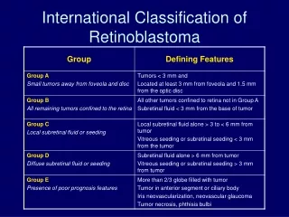
Diagnosis To tell if you have retinoblastoma, your ophthalmologist might do several different procedures that are normally done at any regular check-up. One of the regular procedures is the checking of the eye for dilation of the pupil. Even looking at an eye, you can tell that retinoblastoma is present. The eye will have a yellowish look and appear to be glazed (Figure 1). Some more complex methods of telling, is with a CT scan or even an ultrasound would be appropriate. The tumor can vary in sizes and in fact the size of the tumor will determine what kind of treatment will be used to rid of the tumor (figure 2). Figure 1 Figure 2
Cause The causes of retinoblastoma are mostly genetic with the exception of a rare case of a new case mutation. In fact, the children of a person with the illness has a 50% chance of inheriting the disorder. The whole disease of retinoblastoma is caused by a mutation that occurs because of a cell in the retina that is duplicating numerously and therefore turning cancerous. The retinoblastoma gene copies in a autosomal dominant pattern which means that if even one parent has the gene, the child will have that 50% chance of inheriting retinoblastoma. Even if the gene is given to a child, it does not necessarily mean that cancer is for sure. As shown in the punnett square (Figure 3), there is a 50% chance of retinoblastoma assuming the heterozygous is the retinoblastoma gene. Figure 3
Signs/Symptoms • Poor vision – When retinoblastoma is present, the vision clearness decreases because of the tumor clouding the eye. • Instead of a typical “red” eye in photographs, the eye will appear white – Again, the tumor will switch the iris color and have it appear white in a glazed fashion. • Strabismus – also known as a wandering eye or cross eyes where the eyes appear to be looking around in different directions. • Red eye – due to irritation of the eyes • White spots – There will be white spots that will be floating around the pupil.