Download
1 / 21
210 likes | 315 Views
Chapter 4 Lecture. Chapter 4. The Integumentary System. Introduction. The integumentary system, or integument, is composed of skin, hair, nails, sweat, oil, and mammary glands. Three layers : Epidermis - epithelial tissue Dermis - dense connective tissue proper - irregular

E N D
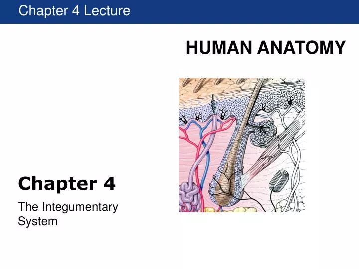
Chapter 4 Lecture Chapter 4 The Integumentary System
Introduction • The integumentary system, or integument, is composed of skin, hair, nails, sweat, oil, and mammary glands. • Three layers: • Epidermis - epithelial tissue • Dermis - dense connective tissue proper - irregular • Hypodermis - loose connective tissue proper and adipose tissue • Functions: • Protection • Thermoregulation • Sense organ • Vitamin D production • Excretion
Integumentary Structure and Function Figure 4.1 Functional Organization of the Integumentary System
Integumentary Structure and Function • Skin, or the cutaneous membrane, has two subdivisions: • Epidermisis the stratified squamous epithelium. • Dermis is the underlying dense irregular CT. • Deep to the dermis is the subcutaneous layer or hypodermis - loose CT and adipose. • Accessory structures include hair, nails, and many multicellular exocrine glands.
Integumentary Structure and Function Figure 4.2 Components of the Integumentary System
The Epidermis • Keratinocytes are the most abundant cells in the epidermis. • At least four different cell layers can be found on most areas of the body. • Melanocytes are pigment cells found deep in the epidermis. • Merkel cells are sensory cells. • Langerhans cells are fixed macrophages.
Layers of the Epidermis Strata of epidermis and the process of keratinization Table 4.1 Epidermal Layers Figure 4.3 Structure of the Epidermis
Thick and Thin Skin Strata granulosum and lucidum Keratohyalin and eleidin precursors to keratin in S. corneum Figure 4.4 Thin and Thick Skin
The Dermis and Subcutaneous Layer Figure 4.2 Components of the Integumentary System
Dermal Organization and the Subcutaneous Layer Figure 4.7 The Structure of the Dermis and the Subcutaneous Layer
Accessory Structures • Hair follicles and hair: • Hair is a nonliving keratinized structure that extends beyond the surface of the skin in most areas of the body. • 98% of the 5 million hairs on the body are not on the head. • Hair follicles are the organs that form the hairs. Figure 4.9a Accessory Structures of the Skin
Glands in the Skin Figure 4.12 A Classification of Exocrine Glands in the Skin
Nails Figure 4.15 Structures of a Nail
Aging and the Integumentary System Figure 4.16 The Skin during the Aging Process
Skin Cancer Types of Skin Cancer: • Actinic Keratosis: Precursor to Squamous Cell Carcinoma • Squamous Cell Carcinoma • Basal Cell Carcinoma • Melanoma • Kaposi's Sarcoma (KS)
Actinic Keratosis • Rough, red or pink scaly patches on sun-exposed areas of the skin, usually <0.5cm in diameter • Precursor lesion for squamous cell carcinoma (Squamous Cell Carcinoma)Up to 1% of these lesions can develop into a Squamous Cell Carcinoma
Squamous Cell Carcinoma • Squamous cell carcinoma is the second most common skin cancer after basal cell carcinoma. • Squamous cell carcinomas are often described as enlarging red bumps, sometimes with a rough, scaly, or crusted surface. They may also look like flat reddish patches in the skin that grow slowly. If untreated, they can become ulcerated (open sores). • Most squamous cell carcinomas grow slowly. • Dull red, rough, scaly raised skin lesions. • Occur most frequently on sun exposed areas (head, neck, ears, lips, back of the hands and forearms) • Sites particularly associated with elevated risk for recurrence or metastasis include: ear, lip and nose
Basal Cell Carcinoma Basal cell carcinomas usually develop on sun-exposed parts of your body, especially your head and neck. A much smaller number occur on the trunk and legs. Yet basal cell carcinomas can also occur on parts of your body that are rarely exposed to sunlight. Although a general warning sign of skin cancer is a sore that won't heal or that repeatedly bleeds and scabs over, basal cell cancer may look like: • A pearly white or waxy bump, often with visible blood vessels on your face, ears or neck. • The bump may bleed, develop a crust or form a depression in the center. In darker skinned people, this type of cancer is usually brown or black. • A flat, scaly, brown or flesh-colored patch on your back or chest. • Over time, these patches can grow quite large.
Melanoma Melanomas can develop anywhere on your body, but they most often develop in areas that have had exposure to the sun, such as your back, legs, arms and face. A-B-C-D-E guide developed by the American Academy of Dermatology: A is for asymmetrical shape. Look for moles with irregular shapes, such as two very different-looking halves B is for irregular border. Look for moles with irregular, notched or scalloped borders — characteristics of melanomas. C is for changes in color. Look for growths that have many colors or an uneven distribution of color. D is for diameter. Look for new growth in a mole larger than about 1/4 inch (6 millimeters). E is for evolving. Look for changes over time, such as a mole that grows in size or that changes color or shape. Moles may also evolve to develop new signs and symptoms, such as new itchiness or bleeding.
Kaposi's Sarcoma (KS) Rare, cancer of the cells that line blood vessels (endothelial cells) • Clinically: brownish-red to blue colored skin lesions found most frequently on legs and feet • Caused by Human Herpes Virus 8 (HHV-8) which causes the cells that line blood vessels (endothelial cells) to become cancerous in the setting of profound and prolonged immunosuppression.