Download
1 / 54
540 likes | 557 Views
Inflammation Lung parenchyma & pleura. Soheir Mahfouz. BRONCHOPNEUMONIA. DEFINITION Patchy suppurative consolidation of the lung centered around a bronchus or bronchiole & involves immediately adjacent alveoli. BRONCHOPNEMONIA --CP. Febrile Extremes of age Cough with productive sputum.

E N D
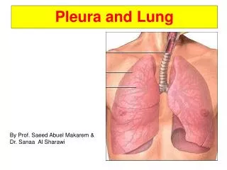
InflammationLung parenchyma & pleura Soheir Mahfouz
BRONCHOPNEUMONIA DEFINITION Patchysuppurative consolidation of the lung centered around a bronchus or bronchiole & involves immediately adjacent alveoli
BRONCHOPNEMONIA --CP • Febrile • Extremes of age • Cough with productive sputum
BRONCHOPNEUMONIA --Etiology Organisms gram +ve cocci: Staph, Strept gram –ve: H. influenza, pseudomonas, E.coli & Klebsiella FOR DIAGNOSIS 1- True sputum sample: diagnostic feature intracellular organisms in neutrophils & macrophages 2- Culture is necessary for determination of organism & antibiotic sensitivities NB In many cases the microbial agent is never diagnosed
Consolidation Patchy, firm & raised
BRONCHOPNEUMONIA –(special presentations) Pseudomonas aeroginosa • Infects lung hematogenously especially in burn patients & immunocompromised. In cystic fibrosis pts. It may be air born and usually fatal • Causes inflammation with necrosis of vascular walls resulting in hemorrhagic pneumonia
Vascular necrosis Hemorrhagic pneumonia
BRONCHOPNEUMONIA –(special presentations) Legionella pneumonia • Neutrophils +manymacrophages in a background of fibrin • Org: short gm –ve coccobacillus DIAGNOSIS: Serology, culture & IF of sputum CP: Acute onset but responds to appropriate antibiotics
HISOPATHOLOGIC VARIANTS OF BRONCHOPNEUMONIA • Neutrophilic or exudative type : common type (SBP) • Histiocytic: Legionella & Mycoplasma • With hyaline membranes: Strept & E. coli • With coagulative necrosis • With abscess formation • With hemorrhage: Pseudomonas (septic vasculitis) & Klebsiella • With leukopenia: in leukemic patients or in chemotherapy patients • With granulomas
Lobar & bronchopneumonia – Complications • Post pneumonic abscess, gangrene • Empyema • Direct spread : bronchopleural fistula & pyopneumothorax • Distant spread: Toxemia, septicemia, pyemia • Lung fibrosis
Interstitial pneumonia & ILD DEFINITION: Inflammation /infection affecting the interstitial tissue , sparing the alveolar spaces NB Atypical pneumonia is a patchy form of interstitial pneumonia & is also called pneumonitis Causes • Viral: respiratory scyncetial virus- cytomegalovirus, HS,Varicella-Zoster,Measles, Adenovirus,Influenza • Mycoplasma • Protozoal: Pneumocystis carnii • Chemical irritants: gases & lipid & aspiration pneumonia • CT diseases: SLE, rheumatoid or mixed
Interstitial pneumonia & Interstitial lung disease (ILD) • Chronic fibrosing pneumonia • Fibrous interstitial pneumonia • Bronchiolitis obliterans organizing pneumonia (BOOP) • Diffuse alveolar damage (DADS)/ AIP • Giant cell interstitial pneumonia • Lymphocytic interstitial pneumonia • Usual interstitial pneumonia • Idiopathic interstitial pneumonia: UIP(IPF)-DIP-NSIP –BOOP(COP) - AIP(ARDS)- RBILD • Pulmonary fibrosis (Diffuse parenchymal lung disease DPLD / ILD) : Pneumonia (UIP – DIP- NSIP) + other causes for lung fibrosis
Acute Interstitial pneumonia (Haman Riche) Chronic Interstitial pneumonia UIP DIP NSIP NB: LIP & GIP are generally secondary pneumonias IDIOPATHIC Interstitial pneumonia NEW Classification
Interstitial pneumonia • UIP usual interstitial pneumonia • Idiopathic interstitial pneumonia • Non specific interstitial pneumonia • Acute necrotizing interstitial pneumonia • LIP lymphocytic interstitial pneumonia
Interstitial pneumonia -Viral • Diffuse interstitial inflammation lymphohistiocytic + edema • More severe cases may have additional changes resembling acute alveolar damage with hyaline membrane formation DIAGNOSIS: LM, serology look for a 4 fold rising titre & DNA probes
OTHER PNEUMONIAS (non infectious) • Aspiration pneumonia • Lipid pneumonia
ASPIRATION PNEUMONIA • Chemical • Bacterial • Mechanical obstruction: fluids & solid objects
Lipid pneumonia • Endogenous : usually associated with Airway obstruction by neoplasm or FB which releases lipids & gives rise to pneumonia • Exogenous :Are a reaction to aspiration of gastric contents or mineral oils gives rise to a FB granulomatous reaction
LIPID PNEUMONIA This is the microscopic appearance of an exogenous lipid pneumonia in which lipid vacuoles appear, mainly along airways, accompanied by an inflammatory response that can contain foreign body giant cells. The term exogenous refers to the origin of the lipid material outside the body. This material is aspirated into the bronchial tree.
LUNG ABSCESS- Types • Aspiration • Post pneumonic • Pyemic Complications of • Bronchiectasis • Infected cysts • Direct spread from liver abscess • Bronchial obstruction
LUNG ABSCESS Chronic Acute
LUNG ABSCESS-pyemic BV Bacteria & pus
LUNG ABSCESS- FATE • SMALL: FIBROSIS • LARGE: CHRONIC ABSCESS • Rupture into a bronchus: hemoptysis,purulent sputum, bronchopleural fistula exposure to saprophytes • Lung gangrene & severe toxemia • Spread by blood: toxemia, septicemia & pyemia • Secondary amyloidosis
Important terms PLEURISY • DEFINITION: Acute fibrinous / serofibrinous inflammation • Definition: collection of fluid in pleural cavity PLEURAL EFFUSIONS
ACUTE NON SUPPURATIVESerofibrinous/Fibrinous Site: Cavities lined by mesothelium • Pleura • Pericardium • Peritoneum • Meninges
Important terms • Terms for abnormal accumulations of fluid: A transudate is protein-poor salt water squeezed through blood vessels by hydrostatic pressure, i.e., it has specific gravity of extracellular fluid, 1.010 or thereabouts. An exudate is an abnormal, protein-rich fluid which has leaked out of inflamed vessels. • A body fluid (either an exudate or an area of liquefaction necrosis) containing neutrophilic leukocytes and necrotic debris is pus. The preferred adjective to describe things with lots of pus is purulent. To produce pus is to suppurate. Pus which literally fills an important body cavity is called an empyema. (This is most common in the pleural cavities.)
Chylothorax The right pleural cavity is filled with a cloudy yellowish-tan fluid, characteristic for a chylothorax. In this case, lymphoma involving the lymphatics of the chest and abdomen led to the collection of chylous fluid. The right lung is markedly atelectatic.
Fibrinous layer Fibrinous layer Submesothelial acute inflammation Submesothelial acute inflammation Congested BV