Download
1 / 52
520 likes | 730 Views
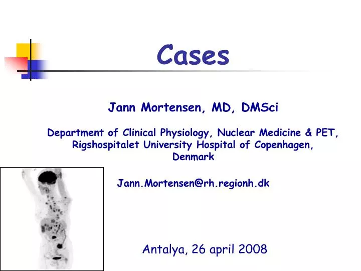
Cases Jann Mortensen, MD, DMSci Department of Clinical Physiology, Nuclear Medicine & PET, Rigshospitalet University Hospital of Copenhagen, Denmark Jann.Mortensen@rh.regionh.dk. Antalya, 26 april 2008.

E N D
Cases Jann Mortensen, MD, DMSci Department of Clinical Physiology, Nuclear Medicine & PET, Rigshospitalet University Hospital of Copenhagen, Denmark Jann.Mortensen@rh.regionh.dk Antalya, 26 april 2008
Case story 166 yrold male, 40 pack-yrs,Presents with coughing and weight lossPrevious history: coronary bypass one year before
Case 1Blood tests, Clinical exam. okLung function by spirometry:FEV1 =2.66 L (96% predicted)
Case 1 • Chest X-ray ? • CT scan ? • PET/CT ? • Bronchoscopy ? • Other ?
Case 1 • Chest X-ray shows • A SPN periperally in left lung (LUL) • A widened mediastinum • Sligth pleural effusion on left side
Case 1 • CT scan ? • PET/CT ? • Bronchoscopy ? • Other ?
Case 1 • Diagnostic PET/CT scan was performed • CT interpretation • PET interpretation • Final PET/CT interpretation
Case 1 • CT scan: • Large tumor mass in left hilum • large lymph nodes bilaterally in Mediastinum • SPN in LUL • Pleural effusion in left side • small lymph nodes in basis of neck
Case 1 • PET and CT both show patology in • tumour in left upper lobe • left hilum • bilaterally in lymph nodes • PET additionally shows • Bone metastases (spine and pelvis) • Patological lymph nodes in basis of neck • Only CT showed pleural effusion
Case 1 • Fiber-bronchoscopy+TBNAB: • Suspicion of tumour in the left upper bronchus • Biopsies inconclusive, SCLC ?
Case 1 • Re-bronchoscopy ? • Re-bronchoscopy with EBUS/EUS? • Mediastinoscopy? • Surgery? • MRI to verify bone metastasis? • Other ?
Case 1 • Re-bronchoscopy with EBUS: • A tumour in 1 segment bronchus in LUL • But biopsy not sufficient • EBUS: shows paratracheal tumour processes bilaterally • TBNAB from the right side, 4R
Case 1 • Final Diagnosis • EBUS TBNAB 4R: SCLC cells • CT: T4 N3 M0 • PET/CT Stage: T4 N3 M1 • Final stage: T4 N3 M1
Case 1 • Final Diagnosis: • SCLC • Extensive disease (T4 N3 M1) • Referred for chemotherapy • Still alive after 6 series, minor regression
New Danish staging algorithm april 2008Fast track and PET/CT • Symptoms (suspicion) -> • max 2 d Chest X-ray, if still suspicion-> • max 2 CT scan, if pot. operable-> • max 2 d PET/CT, d if pot. operable-> • max 2 d Bronchoscopy+TBNAB +/- EBUS/EUS • max 5 d to Patological Diagnosis, if operable-> • max 7 d to Surgery • (Surgery within 14 days from suspicion) • Symptoms (suspicion) -> • max 2 d Chest X-ray, if still suspicion-> • max 2 d PET/CT, if pot. operable-> • max 2 d Bronchoscopy+TBNAB +/- EBUS/EUS • max 5 d to Patological Diagnosis, if operable-> • max 7 d to Surgery • (Surgery within 14 days from suspicion)
Case 2 • 48 yrold male, 20 pack-yrs. • Previous history: colitis ulcerosa • Achalasia for months (CT of abdomen+ EUS performed) • Presents with left sided tonsillar cancer of planocellular type (low grade) Clinical stage T3 N0 M0 • US of neck: no metastases • Plan: MRI (staging) and PET/CT (radiation field plan) • Radiation therapy at Pharyngs+ Neck: 2 Gy * 34 with concommitant Cisplatin
Case 2 : MRI: 17.7.07
Case 2 CT: 20.6.07
Case 2 PET/CT: 24.7.2007
Case 2 PET/CT: 24.7.2007
Case 2 PET/CT: 24.7.2007 • Patologically increased uptake in the • tonsil cancer • lymph nodes in neck bilaterally • 2 lymph nodes in left mediastinum (station 5+6)
Case 2 • Tonsillar metastases ? • Metastasis from tonsillar cancer to the mediastinumare rare. • Another cancer ? • False positive ? (eg. inflammation/infection) • Other ?
Case 2 Do wecare? No, we just include the area in the radiation field Yes, searchcytological/histologicaldiagnosis Other
Case 2 Lung function: FEV1 3.4 L (88% p) DLCO 9.3 (87% p) Postoperative 3.8.07 3.8.07: Bronchoscopy followed by Videoassisted mini-thoracotomy and excision of 2 lymph nodes ”en bloc” from station 5+6 left
Case 2 Patology: Lymph nodes Station 5+6: Low differentiated mucus producing adenocarcinoma, Immunostaining: positive for CK7 + TTF1, surfactant A and CK19 Negative for P63, CK 5/6, thyroglobulin, etc. Thus very likely pulmonary NSCLC
Case 2 Planocellular tonsillar cancer with bilateral neck metastases and potentially curative treatment Metastatic NSCLC (adenocarcinoma) No primary
Case 2 Search for primaryadenocarcinoma ? Wholebody PET/CT negative, repeat ? Repeatesophagealexamination ? Thyroidexamination ? Otherexaminations ? Which first ?
Case 2 • Search for primaryadenocarcinoma • Wholebody PET/CT was negative, repeatYes • Esophagealexamination. CT showedthickening, • But EUS + Biopies: ok, Repeatet: again ok • Thyroidexaminationordered
Case 2 US of thyroid gland 27.11.07: ok
Case 2 2nd PET/CT: 5.10.2007 New 18 mm nodule in RUL with patologically increased uptake (SUV 2.6)
Case 2 Whatnow? Primary NSCLC ? Metastasis? Other ?
Case 2 Wedge resection 24.10.07
Case 2 Patology: Localised fibrosis and organised pneumonia: BOOP Ie ”False positive FDG-PET” Still no primary, what now?
Case 2 4.11.07 Total clinical remission PET/CT control 3 months after radical treatment of tonsil cancer
Case 2 3 rd PET/CT: 12.12.2007 Sequelae after surgery (24.10.2007), no suspicion of malignancy
Case 2 • Dec. 2007 status: Completeremission of • tonsil cancer withneckmetastasis • and metastatic NSCLC (noprimary) • Whatnow? • Follow-op • Chemotherapy for NSCLC
Case 2 4.2.08 Adjuvant chemotherapy for NSCLC with Cisplatin and Vinorelbine