Download
1 / 75
1.3k likes | 6.62k Views
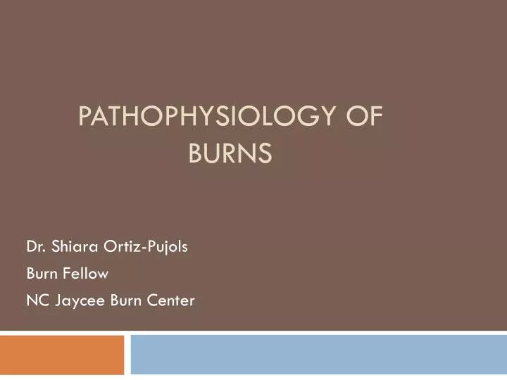
Pathophysiology of Burns. Dr. Shiara Ortiz-Pujols Burn Fellow NC Jaycee Burn Center. Objectives. PART 1 Anatomy Overview Causes of Burns Estimating Burns (Depth & %) Categories & Zones. PART 2 Physiologic Implications Pathophysiology Resuscitation Post-Resuscitation

E N D
Pathophysiology of Burns Dr. Shiara Ortiz-Pujols Burn Fellow NC Jaycee Burn Center
Objectives • PART 1 • Anatomy Overview • Causes of Burns • Estimating Burns (Depth & %) • Categories & Zones • PART 2 • Physiologic Implications • Pathophysiology • Resuscitation • Post-Resuscitation • Board Questions
Anatomy • Adult skin surface 1.5-2.0 m2 (0.2-0.3 in newborns); largest organ • Skin thickness 1-2 mm; peaks age 30-40; M> F • Functions include: • protection from external environment • maintenance of fluid/electrolyte homeostasis • Thermoregulation • immunologic function • sensation • Metabolic organ (i.e., Vit D synthesis)
Causes of Burns • Usually caused by heat, electricity, chemicals, radiation, and friction • Thermal burns are caused by steam, fire, hot objects or hot liquids. • Most common burns for children and the elderly • Electrical burns are the result of direct contact with electricity or lightning • Chemical burns occur when the skin comes in contact with household or industrial chemicals • Radiation burns are caused by over-exposure to the sun, tanning booths, sun lamps, X-rays or radiation from cancer treatments • Friction burns occur when skin rubs against a hard surface, e.g. carpet, gym floor, concrete or a treadmill
Effect of Heat • Temporal and quantitative • 40-44C, enzymes malfunction, proteins denature and pumps fail • > 44C, damage occurs faster than repair mechanisms can keep up with • Damage continues even when the source is withdrawn
Effect of Electricity • Effects of current depend on several factors - Type of circuit - Voltage - Resistance of body - Amperage - Pathway of current - Duration of contact • High voltage (>1000V) causes underlying tissue damage. Deep tissues act as insulators and continue to be injured. • Resistance of various tissues from L→H: nerve, vessels, muscle, skin, tendon, fat, bone Ohm’s Law- V=IR • Damage more related to cross-sectional area which explains extremity injuries without trunk injuries.
Electrical Storms/Lightning • Burns are characteristically superficial and present as a spidery or arborescent pattern. • Cardiopulmonary arrest is common following lightning injury. • Coma and neurologic defects are also common but usually clear in a few hours or days. • Watch for tympanic membrane rupture • Usually lethal in 1/3 of patients. • World record for surviving lightning strikes is Roy C. Sullivan who was a park ranger from VA. Roy was struck 7 times from 1942-1977.
Effect of Chemicals • Acids and alkalis cause injury via different mechanisms. • Petroleum products can cause delipidation and depth of wound 2° tendency to adhere to skin • Acids: • coagulation necrosis • denaturing proteins upon tissue contact • area of coagulation is formed and limits extension of injury • exception is hydrofluoric acid, which produces a liquefaction necrosis similar to alkalis. • Acid damaged skin can look tanned and smooth; do not mistake for a suntan. • Alkalis: • liquefaction necrosis • potentially more dangerous than acid burns • liquefy tissue by denaturation of proteins and saponification of fats • In contrast to acids, whose tissue penetration is limited by the formation of a coagulum, alkalis can continue to penetrate very deeply into tissue • Can cause severe precipitous airway edema or obstruction.
Inhalation Injury • Heat dispersed in upper airways leads to edema • Cooled smoke and toxins carried distally • Increased blood flow to bronchial arteries causes edema • Increased lung neutrophils – mediators of lung damage – release proteases and oxygen free radicals (ROS) • Exudate in upper airways – formation of fibrin casts
Stages of Inhalation Injury • Stage 1 – acute pulmonary insufficiency • Signs of pulmonary failure at presentation • Stage 2 – 72-96 hrs after presentation (ARDS picture) • extravasation of water • Hypoxemia • Lobar infiltrates • Stage 3 – bronchopneumonia • Early – Staph pneumonia (frequently PCN resistant) • Late - Pseudomonas
Inhalation Injury Bronchoscopy: - erythema - intraglottic soot - ulceration
Grading of Burn Wounds • Mild: < 5% TBSA • Moderate: 5-15% TBSA • Severe: > 15% (95% of burns seen) • May require Burn Unit care because of potential for disability despite small TBSA (face, hands, feet, perineum)
Area of Burn – “Rule of 9s” Note that a patient's palm is approximately 1% TBSA and can be used for estimating patchy areas.
Estimation of Burn Wound Depth • Initial assessment is often unreliable • Ignore mild erythema when calculating fluid requirements • Pink areas that blanch are usually superficial • Deeper wounds are dark red, mottled or pale and waxy • Insensate areas are usually deep(3rd degree or greater)
Factors Influencing Wound Depth • Temperature and duration • Thickness of skin (thin on eyelids, thick on back) • Age (children and elderly have proportionally thinner skin in comparison to adults) • Vascularity • Agent – oil vs water; acidic vs alkalotic • Time to definitive care
Burn Zones • Circumferential zones radiating from primarily burned tissues, as follows: • Zone of coagulation - A nonviable area of tissue at the epicenterof the burn • Zone of ischemia or stasis - Surrounding tissues (both deep and peripheral) to the coagulated areas, which are not devitalized initially but, 2° microvascular insult, can progress irreversibly to necrosis over several days if not resuscitated properly • Zone of hyperemia - Peripheraltissues that undergo vasodilatory changes due to neighboring inflammatory mediator release but are not injured thermally and remain viable
Zone of Hyperemia Zone of Ischemia Zone of Coagulation
Categories of Burns – First degree • Burns are divided into 4 categories, depending on the depth of the injury, as follows: • First-degree burns are limited to the epidermis. • A typical sunburn is a first-degree burn. • Painful, but self-limiting. • First-degree burns do not lead to scarring and require only local wound care.
Categories of Burns – Second degree • Second-degree burns • point of injury extends into the dermis, with some residual dermis remaining viable • Partial thickness or Full thickness • those requiring surgery vs those which do not
Categories of Burns – Third degree • Third-degreeor full-thickness burns involve destruction of the entire dermis, leaving only subcutaneous tissue exposed.
Escharatomy Sites Preferred sites for escharotomy incisions. Dotted lines indicate the escharotomy sites. Bold lines indicate areas where caution is required because vascular structures and nerves may be damaged by escharotomy incisions. (From Davis JH, Drucker WR, Foster RS, et al: Clinical Surgery. St. Louis, CV Mosby, 1987.)
Categories of Burns – 4th degree • Fourth-degree burn is usually associated with lethal injury. • Extend beyond the subcutaneous tissue, involving the muscle, fascia, and bone. • Occasionally termed transmural burns, these injuries often are associated with complete transection of an extremity.
PART 2 • Physiologic Implications • Pathophysiology • Resuscitation • Post-Resuscitation • Board Questions
Physiologic Implications of Burn Injury • Predictable changes • Related to period of injury • Can be anticipated
Pathophysiology of Burns • Cell damage and death causes vasoactive mediator release: • Histamines • Thromboxanes • Cytokines • Increasing capillary permeability causes edema, third spacing and dehydration • Possible obstruction to circulation (compartment syndrome) and/or airway
Resuscitation Period • “early ebb with late flow”; days 0-3 • Hypodynamic, with need for close fluid resuscitationmonitoring • Massive, diffuse capillary leak2° to inflammatory mediators; abates 18-24 hrsafter injury and volume requirements abruptly decline • leak can be seen in those with delayed resuscitation 2° systemic release of O2 radicals upon reperfusion • Extravascular extravasation of fluid, lytes, colloid molecules • Other variables affect resuscitation: preexisting fluid deficits, delay until treatment, inhalation injury, depth of wound • Must reevaluate resuscitation progress and endpoints frequently; do not just use a formula
Postresuscitation Period • Day 3 until 95% wound closure • Hyperdynamic, febrile, protein catabolic state • Tachycardia can be normal in burn patients • Blood pressure may be hard to obtain due to circumferential burns • Release of more inflammatory mediators, cortisol, glucagon, catecholamines, bacteria from wound • High risk of infection and pain • Remove non-viable tissue or close wounds to avoid sepsis • Nutritional support essential • Maintain and support body temperature with high ambient temps and humidity
Recovery Period • 95% wound closure until 1 year post-injury • Continued catabolism and risk of non-healing wound • Anticipate septic events, treat complications, and continue nutritional support
Pathophysiology of Electrical Burns • Small cutaneous lesions may overlie extensive areas of damaged muscle → myoglobin ARF. • Monitor for at least 48 hours after injury for cardiopulmonary arrest • May see vertebral compression fracturesfrom tetanic contractions or other fractures from a fall. • Visceral injury is rare but liver necrosis, GI perforation, focal pancreatic necrosis and gallbladder necrosis have been reported. • Look for motor and sensory deficits—motor nerves are affected more than sensory nerves. • Thrombosis of nutrient vessels of the nerve trunks or spinal cord can cause late onset deficits. Early deficits are direct neuronal injury. • Delayed hemorrhage can occur from affected vessels • Cataracts may form up to 3 or more years after electrical injury • Microwave radiation damages tissues via a heating effect. Subcutaneous fatty tissue is often spared given its lower water content.
Burn Edema and Inflammation • Generalized edema found in burns > 30% TBSA • Heat directly damages vessels and causes permeability • Heat activates complement histamine release and more permeability thrombosis and coagulation systems +
Systemic Response to Burn Injury • Accelerated fluid loss 2° leaky capillaries • Host resistance to infection • Multisystem Organ Failure • Infections in burns <20% TBSA are well tolerated. • > 40% TBSA with infection has very low survival rate • Initially CO, subsequent hypermetabolic state w/ doubling of CO in 24 – 48 hours