Download
1 / 42
430 likes | 439 Views
Innate Immunity- First Line of Defense. What is the Innate Immune System?. includes physical, chemical, and cellular barriers physical barriers include skin and mucus membranes chemical barriers include stomach acidity, secreted anti-microbial peptides

E N D
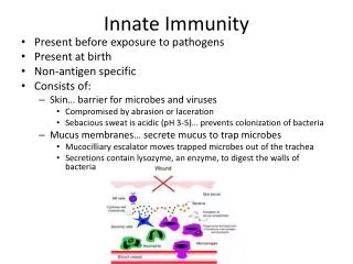
What is the Innate Immune System? • includes physical, chemical, and cellular barriers • physical barriers include skin and mucus membranes • chemical barriers include stomach acidity, secreted anti-microbial peptides • cellular barriers include macrophages, neutrophils • innate immune response activation occurs within minutes of pathogen recognition
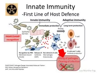
Epithelial defense mechanisms Examples of chemical barriers: - lysozyme, phospholypase A (saliva, tears) - acid pH (stomach) - anti-fungal peptides called alpha-defensins (intestinal tract) - anti-microbial peptides called beta-defensins (respiratory, urogenital tract) - surfactant-A and -D proteins opsonize pathogens for enhanced phagocytosis (lung) Additional nonspecific defense mechanism is endogenous commensial (non-pathogenic) bacterial flora (microbiota)
2-2 Epithelial surfaces of the body provide the first barrier against infection
2-2 Epithelial surfaces of the body provide the first barrier against infection
What happens when the physical and chemical barriers are breached?
Innate Immunity- First Line of Defense • Characteristics: • rapid • does not generate immunologic memory • dependent upon germline encoded receptors recognizing structures common to many pathogens Innate Immunity
Innate Immune Receptors • Innate immune receptors are not clonally distributed • Binding of receptors results in rapid response • Innate immune receptors mediate three functions: - phagocytic receptors to stimulate pathogen uptake - chemotactic receptors that guide phagocytes to site of infection - stimulate production of effector molecules and cytokines that induce innate responses and also influence downstream adaptive immune responses
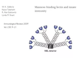
Pathogen Recognition • Most microorganisms express repeating patterns of molecular structures termed Pathogen Associated Molecular Patterns (PAMPs) • Innate immune system has evolved mechanisms capable of recognizing these repeating patterns termed Pattern Recognition Receptors (PRRs) • Examples of Pattern Recognition Receptors: - Mannose-Binding Lectin (MBL) - Macrophage Mannose Receptor - Scavenger Receptors - Toll-like Receptors (TLRs) - Nod-like Receptors (NLRs) - RNA helicases (RIG-I, MDA-5)
Toll-Like Receptors (TLRs) TNFa IFNab Cellular Localization: • Lysosomal localization (i.e. subcellular) of TLR-3 and TLR7-9 • TLR-3 and 7-9 recognize viral/bacterial nucleic acids • lysosomal expression isolates pathogen nucleic acid recognition away from potential cross-reaction with host mammalian nucleic acid motifs
Cells also have cytoplasmic receptors to help sense viral nucleic acid • RIG-I Is a cytosoIic protein that detects viral RNA. • 5'-trIphosphate and double-stranded (ds) RNA are two molecular patterns that enable RIG-I to discriminate pathogenic from self-RNA. • Two N-terminal caspase activation and recruitment domains (CARDs) transmit the signal and the regulatory domain prevents signaling in the absence of viral RNA. • MDA-5 works similarly Induce IFNab
NLRs are cytoplasmic bacterial sensors that activate IL-1b “inflammasomes” Mariathasan and Monack Nature Reviews Immunology7, 31–40 (January 2007) | doi:10.1038/nri1997
Inflammatory Cytokines • Activation of TLRs, NLRs and RIG-I type molecules results in production of TNFa, IFNab and IL-1b • These cytokines are critical for host defense • TNFa activates macrophage and PMN phagocytosis and killing • IFNab activates anti-viral mechanisms • IL-1 stimulates inflammation and fever
What happens next? • If a pathogen is detected by a phagocytic cell type (macrophages and PMNs), the phagocytic cell will attempt to destroy the infection
Macrophage Microbial Killing Once the PRRs are activated by the PAMPs, phagocytosis is initiated • Phagocytosis is active process: • - Internalization of pathogen into phagosome • - Acidification of phagosome • Fusion of phagosome with lysosomes that contain anti-microbial compounds (phagolysosome) • This may be sufficient to kill the pathogen • If not, reactive oxygen and nitrogen species may need to be generated
Generation of Antimicrobial Species in macrophages and PMNs L-arginine + O2 + NADPH NO + L-citrulline + NADP Inducible nitric oxide synthetase (iNOS)
Chronic Granulomatous Disease (CGD): - caused by mutation in NADPH oxidase enzyme complex - most common form is X-linked - heightened susceptibility to infections, particularly intracellular bacteria - form granulomas due to inability to kill phagocytosed bacteria
Activated macrophages secrete proteins that drive innate response Cytokines - induce response by binding to specific receptors - can function in autocrine or paracrine manner - cytokines (and their receptors) are clustered according to structural similarities - critical cytokines secreted by macrophages following activation include TNFa, IL-1, IL-6, IL-12 to stimulate inflammation and phagocytosis/killing • Chemokines • - diverse family of chemotactic cytokines, induce directed chemotaxis of cells • all related in amino acid structure • - certain chemokines induce cell activation in addition to cell recruitment • - promiscuous in receptor usage, each can bind more than one receptor • - likewise, receptors are promiscuous
Clinical symptoms of inflammation: pain, redness, heat, swelling 1. Increased vascular diameter, increased blood flow (heat, redness) 2. Activation of vascular endothelium to express adhesion molecules, increases leukocyte binding 3. PMNs are first cell type recruited to site, followed later by monocytes 4. Increased vascular permeability results in local swelling and pain Microvascular coagulation helps prevent pathogen spread into bloodstream (physical barrier)
Chemokines • Infection induces the release of various chemokines • Theses substances bind specific and sometimes shared receptors to recruit various types of immune cells to the site of infection
Viruses induce Interferon-a/b production - Termed interferons because they “interfere” with viral replication. - IFN-a/b produced by many diverse cell types following viral infection. - Synthesized in response to dsRNA that is not found in mammalian cells. - TLR-3, RIG-1, MDA-5 all recognize dsRNA. • IFN-a/b actions for viral defense: • - secreted IFNs bind to cell surface IFN-receptor in autocrine and paracrine fashion • - induce host cell proteins that inhibit viral replication • - enhance cellular immune responses against virus • - upregulate MHC class I molecules • active Natural Killer (NK) cells to lyse infected cells and to secrete cytokines • IFNs serve as a firebreak to prevent spread of virus in tissue
Natural Killer (NK) Cells • First identified by having the ability to lytically kill certain tumor cell lines without prior sensitization • Kill target cell by release of cytotoxic granules containing granzymes and perforin which penetrate target cell membrane and induce programmed cell death • Can mediate Antibody-Dependent Cellular Cytotoxicity (ADCC) • Kill virally-infected cells with missing MHC class I • Activated by IFN-a/b or IL-12 (produced rapidly by activated macrophages) • Activated NK cells secrete IFNg, acts on macrophages to increase microbial phagocytosis and killing
NK cell receptors Inhibitory Receptors: - germ-line encoded, no combinatorial diversity as seen with T/B cell receptors - inhibit cytotoxicity to prevent killing of normal host cells - specific for MHC class I alleles - binding to class I sends inhibitory signal to NK cells Activating Receptors: - germ-line encoded - recognize carbohydrate structures on self proteins
NK Cell Cytotoxicity • Schematic representation of NK cell receptors and killing. Normal cells are not killed because inhibitory signals from MHC class I molecules override activating signals. In tumor cells or virus-infected cells, reduced expression or alteration of MHC molecules interrupts the inhibitory signals, allowing activation of NK cells and lysis of target cells. Virus-infected or
“Innate” Lymphocytes • Unique, minor subsets of T and B lymphocytes that undergo receptor gene rearrangements to generate receptor diversity (unlike NK cells) • These subsets express limited receptor diversity, utilizing only a small number of receptor gene segments • - Tend to found in specific locations in the body, usually sites that encounter exogenous antigens or pathogens
B-1 (CD5) B Lymphocytes Distinguished from conventional B cells by expression of CD5 Likely to by the B-cell equivalent of gd-T cells (arise very early in ontogeny, limited and distinctive gene rearrangements) Found in distinct microenvironment (peritoneal cavity and pleural spaces) Secrete IgM antibodies without need of T cell help (unlike conventional B cells), results in rapid response (within 48 hours); termed natural antibodies Antibody responses to bacterial polysaccharide components of cell wall No immunological memory generated
Gamma-Delta T cells Generated very early in ontogeny (prior to birth). Represent the first T cell subsets generated in the thymus Two subsets: - one subset utilizes diverse gd T cell receptor rearrangements and are found in all lymphoid tissues - one utilizes TCR of very limited diversity. Found in high numbers in mucosal linings (pulmonary, urogenital, gastrointestinal) and skin, termed intraepithelialgd T cells T cell receptor ligands: - self-proteins expressed by damaged, injured, stressed epithelium (heat shock proteins, unique MHC-associated molecules, phospholipids) - products of bacterial metabolism and breakdown (small organic phosphates, alkylamines)
Ag recognition by gd-T cells usually does not require MHC presentation, occurs directly (similar to antibody); detailed 3-D structure ofgd-TCR more closely resembles antibody than ab-TCR Direct recognition of TCR-ligand without requirement of antigen processing allows for rapid response
Natural Killer-T Cells (NK-T Cells) • - Minor subset of ab-T cells originally described by expression of NK-cell associated markers • - Majority express invariant TCR (V14-J18/V8.2), remaining express diverse TCR • - Rapidly release large amounts of IL-4 and IFNg, can interact with/influence other “innate immune” lymphocytes (NK cells, gd-T cells) • - Recognize self and foreign glycolipids presented by CD1 • - Crystal structure analysis of CD1d indicates the presence of an MHC-like fold with a large, hydrophobic binding groove • - Due to the unique glycolipid antigen binding ability of CD1 molecules, it has been speculated that CD1 acts as an alternative mechanism for surveillance of foreign and altered-self glycolipids that would otherwise escape conventional class I and II pathways
Dendritic Cells • DCs link innate and adaptive immunity • DCs are immature as they circulate waiting to encounter pathogens • At this point, they are highly phagocytic, but not good stimulators of adaptive T cell responses • Once they are activated by pathogens and activation of their PRRs, they secrete cytokines to initiate inflammation and then they migrate to lymph nodes and mature • As mature DCs they are excellent APCs for T cell stimulation
DC migration and antigen presentation: interface between innate and adaptive immunity Immature DC - principal function is antigen capture - highly phagocytic - low T cell stimulating potential low MHC class II low CD80/86 expression Mature DC - principal function is antigen presentation - low phagocytic capacity - high T cell stimulating potential high MHC class II high CD80/86 expression
A view of DC-T cell interactions DC: Note dendrites
Complement • There is a system of >30 proteins that collectively make up the complement system (originally named because they complement Ab functions) • These proteins can recognize some pathogen surfaces intrinsically, or can recognize Ab molecules bound to pathogen-infected cells • The recognition by and activation of the complement system results in pore-forming complexes being created on infected cells which results in lysis of infected cells • Other byproducts of complement activation are the recruitment of more immune effector cells by the C3a and C5a components • There are classical and alternative pathways of complement activation, but they lead to the same outcome
Ab-mediated Not Ab mediated
Figure 7.11: Complement cascade pathways. Adapted from S. Smith. “Immunologic Aspects of Organ Transplantation.” Organ Transplant. Medscape, 2002.