Download
1 / 33
330 likes | 556 Views
Objective 6. Show the major regions of the brain and describe their functions. . Regions of the Brain. Cerebral hemispheres Diencephalon Brain stem Cerebellum. Figure 7.12b. Cerebral Hemispheres (Cerebrum). Paired (left and right) superior parts of the brain

E N D
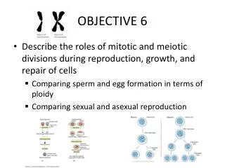
Objective 6 Show the major regions of the brain and describe their functions.
Regions of the Brain • Cerebral hemispheres • Diencephalon • Brain stem • Cerebellum Figure 7.12b
Cerebral Hemispheres (Cerebrum) • Paired (left and right) superior parts of the brain • Include more than half of the brain mass Figure 7.13a
Cerebral Hemispheres (Cerebrum) • The surface is made of ridges (gyri) and grooves (sulci) Figure 7.13a
Lobes of the Cerebrum • Fissures (deep grooves) divide the cerebrum into lobes • Surface lobes of the cerebrum • Frontal lobe • Parietal lobe • Occipital lobe • Temporal lobe
Lobes of the Cerebrum Figure 7.15a
Specialized Areas of the Cerebrum • Somatic sensory area – receives impulses from the body’s sensory receptors • Primary motor area – sends impulses to skeletal muscles • Broca’s area – involved in our ability to speak
Sensory and Motor Areas of the Cerebral Cortex Figure 7.14
Specialized Functions of the Cerebrum • Cerebral areas involved in special senses • Gustatory area (taste) • Visual area • Auditory area (hearing) • Olfactory area (smell) • Interpretation areas of the cerebrum • Speech/language region • Language comprehension region • General interpretation area
Specialized Areas of the Cerebrum Figure 7.13c
Layers of the Cerebrum • Gray matter • Outer layer • Composed mostly of neuron cell bodies Figure 7.13a
Layers of the Cerebrum • White matter • Fiber tracts inside the gray matter • Example: corpus callosum connects hemispheres Figure 7.13a
Diencephalon • Sits on top of the brain stem • Enclosed by the cerebral hemispheres • Made of three parts • Thalamus • Hypothalamus • Epithalamus
Diencephalon Figure 7.15
Thalamus • The relay station for sensory impulses • Transfers impulses to the correct part of the cortex for localization and interpretation
Hypothalamus • “Under” the thalamus • Important autonomic nervous system center • Helps regulate body temperature • Controls water balance • Regulates metabolism • An important part of the limbic system (emotions)
Brain Stem • Attaches to the spinal cord • Parts of the brain stem • Midbrain • Pons • Medulla oblongata
Brain Stem Figure 7.15a
Midbrain • Has two bulging fiber tracts – cerebral peduncles • Has four rounded protrusions – corpora quadrigemina • Reflex centers for vision and hearing
Pons • The bulging center part of the brain stem • Includes nuclei involved in the control of breathing
Medulla Oblongata • The lowest part of the brain stem • Merges into the spinal cord • Contains control centers for • Heart rate control • Blood pressure regulation • Breathing • Swallowing • Vomiting
Cerebellum • Two hemispheres with convoluted surfaces • Provides involuntary coordination of body movements
Cerebellum Figure 7.15a
Objective 7 Describe how meninges, cerebrospinal fluid, and the blood brain barrier protect the Central Nervous System.
Protection of the Central Nervous System • Scalp and skin…..Skull and vertebral column • Meninges Cerebrospinal fluid • Blood brain barrier Figure 7.16a
Meninges • Dura mater • Double-layered external covering • Periosteum – attached to surface of the skull • Meningeal layer – outer covering of the brain • Folds inward in several areas
Meninges • Arachnoid layer • Middle layer • Web-like • Pia mater • Internal layer • Clings to the surface of the brain
Cerebrospinal Fluid • Similar to blood plasma composition • Formed by the choroid plexus • Forms a watery cushion to protect the brain • Circulated in arachnoid space, ventricles, and central canal of the spinal cord
Ventricles and Location of the Cerebrospinal Fluid Figure 7.17a–b
Ventricles and Location of the Cerebrospinal Fluid Figure 7.17c
Blood Brain Barrier • Includes the least permeable capillaries of the body • However many substances (some potentially harmful) can pass through… • Fats and fat soluble molecules • Respiratory gases • Alcohol • Nicotine • Anesthesia
Traumatic Brain Injuries • Concussion • Slight brain injury • No permanent brain damage • Contusion • Nervous tissue destruction occurs • Nervous tissue does not regenerate • Cerebral edema • Swelling from the inflammatory response • May compress and kill brain tissue