Download
1 / 25
310 likes | 839 Views
HEMOPOIESIS. Dr. Dalia Kamal Eldien. What is Hemopoiesis?.

E N D
HEMOPOIESIS Dr. Dalia KamalEldien
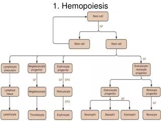
What is Hemopoiesis? • Hematopoiesis refers to the formation and development of all types of blood cells from their parental precursors. In postnatal life in humans, erythrocytes, granulocytes, monocytes, and platelets are normally produced only in the bone marrow. Lymphocytes are produced in the secondary lymphoid organs, as well as in the bone marrow and thymus gland. The main blood cell groups including the red blood cells, white blood cells and platelets are derived from a multipotent stem cell.
Formation of Red Blood Cells • Erythropoiesis is the formation of erythrocytesfrom the progenitor cells through a process of mitotic growth and maturation. The first recognizable erythyroid cell in the bone marrow is the proerythroblast or pronormoblast, which on Wright or Giemsa stain is a large cell with basophilic cytoplasm and an immature nuclear chromatin pattern. Subsequent cell divisions give rise to basophilic, polychromatophilic, and finally orthochromatophilic normoblasts, which are no longer capable of mitosis.
Reticulocytes remain within the bone marrow for approximately 2 days as they continue to accumulate hemoglobin and lose some of their RNA. The reticulocyte then enters the peripheral blood, were, after about one more day, it loses its residual RNA and some of its excessive plasma membrane and becomes indistinguishable form adult erythrocytes. Under normal conditions the transit time from the pronormoblast to the reticulocyte entering the peripheral blood is about 5 days.
Normal morphology of RBCs • Red blood cells have a funny shape – they are a biconcave disc, that is, they look kind of like a flat donut but without the hope in the middle. They are very flexible and can change shape very easily so they can squeeze through your very tiny capillaries.
Alterations in Erythrocyte Shape • Spherocyte. • Stomatocyte. • Schizocyte. • Megalocyte. • Poikilocyte. • Leptocyte. • Dacrocyte. • Acanthocyte. • Ovalocyte. • Rouleaux Formation.
Formation of White Blood Cells • Neutrophils and monocytes, which evolve into macrophages when they enter the tissues, are arise form a common progenitor. The myeloblast is the earliest recognizable precursor in the granulocytic series that is found in the bone marrow. On division the myeloblast gives rise to promyelocyte which contain abundant dark primary granules that overlie both nucleus and cytoplasm.
This cell division and maturation sequence continues form promyelocytes to early myelocytes, late myelocytes, and then metamyelocytes, which are no longer capable of cell division. As the metamyelocyte matures the nucleus becomes more attenuated and the cell is then called a (band) or (stab) form. Subsequent segmentation of the nucleus gives rise to the matureneutrophil or polymorphonuclear leucocyte.
The average interval from the initiation of granulopoiesis to the entry of the mature neutrophil into the circulation is 10 to 13 days. The mature neutrophil remains in the circulation for only about 10 to 14 hours before entering the tissue, where it soon dies after performing its phagocytic function. • The early maturation of the basophilic and eosinophilic granulocyte is similar to that of the neutrophlic granulocyte.
Since the monoblast can not be differentiated from the myeloblast on morphologic or histochemical criteria, one may assume that the myeloblast can give rise to myeloid monocytic cells.
Monoblsat • Promonocyte • Monocyte • Macrophage
Lymphopoiesis • The precursor of the lymphocyte is believed to be the primitive multipotent stem cell that also gives rise to the multipotent myeloid stem cell for the granulocytic, erythyroid, and megakaryocytic cell lines. • Lymphoid precursor cells travel to specific sites, where they differentiate into cells capable of either expressing cell- mediated immune responses or secreting immunoglobulins.
The influence for the former type of differentiation in humans is the thymus gland; the resulting cells are defined as thymus-dependent lymphocytes, or T cells. • The site of the formation of lymphocytes with the potential to differentiate into antibody-producing cells has not been identified in humans, although it may be the tonsils or bone marrow. B cells ultimately differentiate into antibody-producing cells called plasma cells.
Formation of Platelets • Platelets are produced in the bone marrow by fragmentation of the cytoplasm of megakaryocytes. The precursor of the megakaryocyte is the megakaryoblast. The nuclear DNA of megakaryoblasts and early megakaryocytes reduplicates without cell division, a process known as endomitosis. • It takes approximately 5 days from a megakaryoblast to become a mature megakaryocyte. Each megakaryocyte produces from 1000 to 8000 platelets. The platelet normally survives form 7 to 10 days in the peripheral blood.
Hematopoietic Regulatory Factors • Stem Cell Growth Factor (Steel factor):Stimulates multipotent hematopoietic stem cells (hemocytoblasts). • Interleukin-3 (multi-CSF*): Stimulates multipotent hematopoietic stem cells and progenitors of eosinophils, neutrophils, basophils, monocytes, and platelets. • Granulocyte-Macrophage CSF (GM-CSF): Stimulates development of erythrocytes, platelets, granulocytes, and monocytes.
Macrophage CSF (M-CSF): Stimulates development of monocytes and macrophages. • Granulocyte CSF (G-CSF): Stimulates development of neutrophils. • Interleukin-5: Stimulates development of eosinophils. • Interleukin-7: Stimulates development of B – lymphocytes. *CSF=Colony stimulating factor
Extramedullary Hemopoiesis • The formation of normal blood cells outside of the bone marrow mainly in the liver and spleen in post fetal life is known as Extramedullary Hemopoiesis.