Download
1 / 10
100 likes | 107 Views
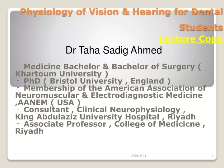
Physiology of Vision & Hearing for Dental Students Lecture Copy. Dr Taha Sadig Ahmed. Medicine Bachelor & Bachelor of Surgery ( Khartoum University ) PhD ( Bristol University , England ) Membership of the American Association of Neuromuscular & Electrodiagnostic Medicine ,AANEM ( USA )

E N D
Physiology of Vision & Hearing for Dental StudentsLecture Copy Dr Taha Sadig Ahmed Medicine Bachelor & Bachelor of Surgery ( Khartoum University ) PhD ( Bristol University , England ) Membership of the American Association of Neuromuscular & Electrodiagnostic Medicine ,AANEM ( USA ) Consultant , Clinical Neurophysiology , King Abdulaziz University Hospital , Riyadh Associate Professor , College of Medicicne , Riyadh
Starts from Pinna ( auricle ) which is the external part , and ends at Tympanic Membrane . the Pinnacollects sound waves ( like a funnel ) and passes them into the External Auditory Meatus the External Auditory meatus transmits these sound waves to cause vibrations in the Tympanic Membrane . these vibrationa are transmitted to the Middle Ear . A/ ExternaL Ear
B/ Middle Ear It is an air-filled cavity . Lies between Tympanic membrane and Oval and Round windows Communicates with pharynx via Eustachean tube Contains 3 auditory ossicles(bones) , called Malleus , Incus & Stapes . Contains tensor tympani and stapediusmuscles .
The Middle Ear ( Contd ) Figure 17.21
Membranous labyrinth contains endolymph Bony labyrinth surrounds and protects membranous labyrinth Cochlea Cochlea القوقعةcontains the receptors for hearing C/ Inner Ear
The Organ of Corti is the receptor for sound • The Cochlea contains a fluid called Perilymph . • The Organ of Corti inside the Cochlea is the receptor for sound . • It is located ( resting ) on the Basilar membrane • Contains inner and outer hair cells . • Extends from the base to the apex of the • It contains hair cells which , when they vibrate , produce Action Potentials in the 8th Cranial Nerve ( Cochlear nerve ) .
Sound Pathway • Sound waves travel toward tympanic membrane, which vibrates • Auditory ossicles conduct the vibration into the inner ear • Movement at the oval window applies pressure to the perilymph of the cochlea • Pressure waves move hair cells in the Organ of Corti • This leads to generation of Action Potentials in the Cochlear nerve . • Cell-bodies of the Cochlear nerve are located in the spiral ganglion of the cochlea • Their afferent fibers synapse in the Cochlear Nucleus in the medulla Inferior Colliculus Medial Geniculate Body Temporal Lobe we hear sound .
You should now be familiar with: • The sensory organs of smell, and the olfactory pathways in the brain. • The accessory and internal structures of the eye, and their functions. • How light stimulates the production of nerve impulses, and the visual pathways. • The structures of the external and middle ear and how they function. • The parts of the inner ear and their roles in hearing. • The pathways for the sensation of hearing.