Download
1 / 20
200 likes | 324 Views
Fabrication of an electrospun nanofibrous scaffold for use in the field of tissue engineering. Tyler Crawford Shannon Daily. Purpose:.

E N D
Fabrication of an electrospun nanofibrous scaffold for use in the field of tissue engineering Tyler Crawford Shannon Daily
Purpose: To create a polycaprolactone mesh which enables cell activity and seeks to eventually provide an application in the field of tissue engineering toward biomimetic skin graft.
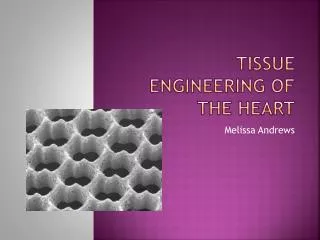
The Extracellular Matrix (ECM) • ECM - main structural tissue of skin • Helps skin renew and generate • Provides signals to intercellular pathways • Engineered ECMs are known as scaffolds
Electrospinning • Ability to create scaffolds • Mimic the ECM (size and porosity) • High surface to volume ratio • Easy to vary mechanical and biological properties through changing materials • Flexible- allows cells to manipulate their environment
Polycaprolactone (PCL) • Biocompatible polymer • Biodegradable at a rate that allows increased cell growth and stability • Easy to manipulate • Relatively low melting point - easy to use
Polycaprolactone • Clinically safe (FDA approval) • Proven to have potential for scaffolds in relation to tissue regeneration • Has created scaffolds w/ ideal conditions • High porosities • Large amounts of surface areas
Additional Biochemical Material • Adding another biochemical can: • Increase stress resistance • Provide better adhesion of cells to the final scaffold • Increase the potential for cell proliferation • Biochemical should • Be a component of skin naturally • Must be able to be combined in a solution to be electrospun
Chitosan (CHT) • Natural polymer that exhibits biocompatible and biodegradable qualities • Cellular binding capabilities • Anti-bacterial properties • High viscosity which limits electrospinning
Experimental Design Procedure: • Create control meshes of pure PCL • Solution= PCL and acetic acid (solvent) • Electrospin • Starting parameters: 15 wt.% concentration, 20 cm from tip of syringe to collector plate, & 20 kV
Procedure continued: • Vary voltage to create 9 meshes • 3 Voltages- 3 trials for each • 20 kV • 15 kV • 25 kV • Examine mesh using Scanning Electron Microscope (SEM) • Culture fibroblast cells onto mesh
Procedure continued: • Observing cells • Inverted light microscope • Analyze cell growth • Cell counts in cells per unit area (mm2) • Means and standard deviations • ANOVA (Analysis of Variance) tests
Procedure continued: • Create solutions of PCL and chitosan • Electrospin • Vary concentration of chitosan to PCL • .5% CHT • 1% CHT • 2% CHT • Total of 9 meshes (3 trials of each concentration)
Procedure continued: • Analyze with SEM • Culture fibroblast cells and seed into meshes created • Determine cell density • Analyze with means, standard deviations, and ANOVA tests
Data and Analysis: • Data obtained: • Fiber diameter and pore diameter of mesh • Cell density amounts • Analysis includes: • Means* • Standard Deviations* • ANOVA tests • 3 comparisons *5-7 measurements/areas for these methods
Data continued: • 15 wt.% solution created • 17 g. acetic acid, 3 g. PCL • Electrospun • 5 mL syringe with bevel tip • Flow rate: .02?? • Mesh created within 2 hrs.
Progress • Background Research • Experimental Design • ISEF (International Science and Engineering Fair) Forms • Started solutions • Just began spinning
References • Akhyari, P., Kamiya, H., Haverich, A., Karck, M., & Lichtenberg, A. (2008). Myocardial tissue engineering: The extracellular matrix. European Journal of Cardio-Thoracic Surgery, 34, 229-241. doi: 10.1016/j.ejcts.2008.03.062 • Bhardwaj, N. & Kundu, S. C. (2010). Electrospinning: A fascinating fiber fabrication technique. Biotechnology Advances, 28, 325-347. doi: 10.1016/j.biotechadv.2010.01.004 • Chong, E.J., Phan, T.T., Lim, I.J., Zhang, Y.Z., Bay, B.H., Ramakrishna, S., & Lim, C.T. (2007). Evaluation of electrospun PCL/gelatin nanofibrous scaffold for wound healing and layered dermal reconstitution. Acta Biomaterialia, 3, 321-330. doi: 10.1016/j.actbio.2007.01.002 • Geng, X., Kwon, O-H., & Jang, J. (2005). Electrospinning of chitosan dissolved in concentrated acetic acid solution. Biomaterials, 26, 5427-5432. • Han, J., Branford-White, C.J., & Zhu, L.M. (2010). Preparation of poly(є-caprolactone)/poly(trimethylene carbonate) blend nanofibers by electrospinning. Carbohydrate Polymers, 79, 214-218. doi: 10.1016/j.carbpol.2009.07.052 • Homayoni, H., Ravandi, S.A.H., & Valizadeh, M. (2009). Electrospinning of chitosan nanofibers: Processing optimization. Carbohydrate Polymers, 77, 656-661. • Lowery, J.L., Datta, N., & Rutledge, G.C. (2010). Effect of fiber diameter, pore size and seeding method on growth of human dermal fibroblasts in electrospun poly(є-caprolactone) fibrous mats. Biomaterials, 31, 491-504. doi: 10.1016/j.biomaterials.2009.09.072 • Nisbet, D.R., Forsythe, J.S., Shen, W., Finkelstein, D.I., & Horne, M.K. (2009). A review of the cellular response on electrospun nanofibers for tissue engineering. Journal of Biomaterials Application, 24, 7-29. • Pham, Q.P., Sharama, V., & Mikos, A.G. (2006). Electrospinning of polymeric nanofibers for tissue engineering applications: A review. Tissue Engineering, 12,1197-1211. • Shevchenko, R.V., James, S.L., & James, S.E. (2010). A review of tissue-engineered skin bioconstructs available for skin reconstruction. Journal of the Royal Society Interface, 7, 229-258. doi: 10.1098/rsif.2009.0403 • Sill, T.J., & von Recum, H.A. (2008). Electrospinning: Applications in drug delivery and tissue engineering. Biomaterials, 29, 1989-2006. doi: 10.1016/j.biomaterials.2008.01.011 • Woodruff, M.A., & Hutmacher, D.W. (in press). The return of a forgotten polymer- Polycaprolactone in the 21st century. Progress in Polymer Science. doi: 10.1016/j.progpolymsci.2010.04.002
THE END ANY QUESTIONS?