Download
1 / 12
120 likes | 140 Views
Hysteroscopy is a procedure used to view the inside of the uterus through a telescope-like device called a hysteroscope. Hysteroscopy offers a valuable extension to the gynecologistu2019s armamentarium.

E N D
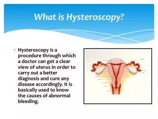
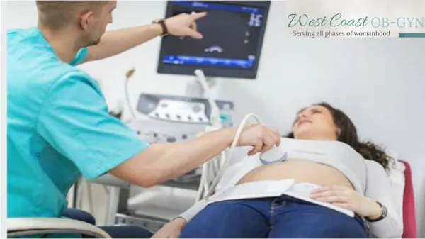
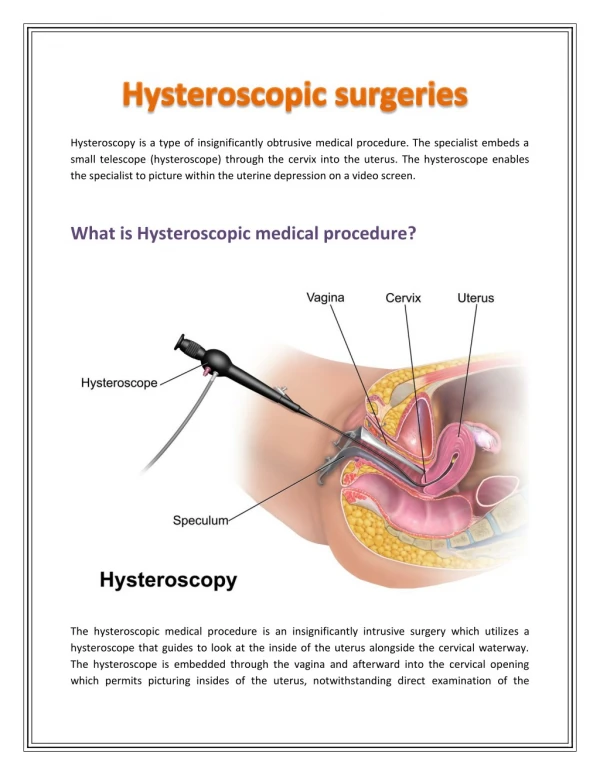
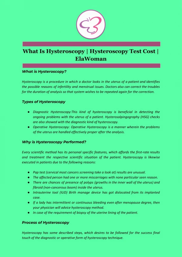
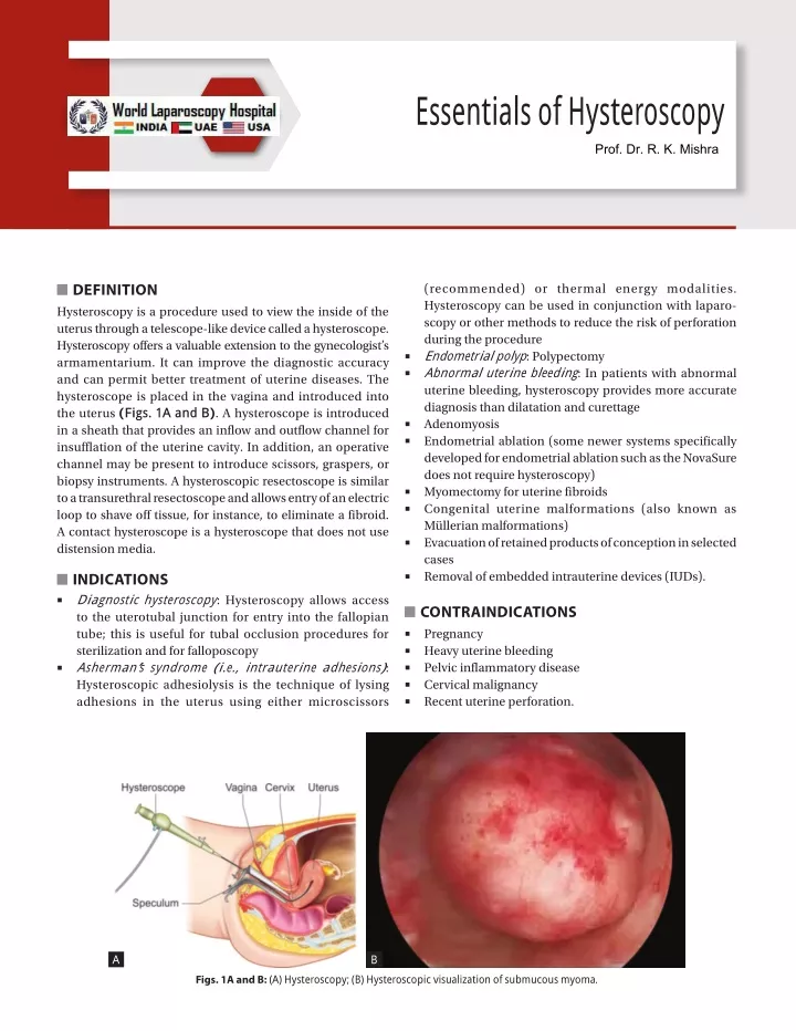
Essentials of H ysteroscopy Prof. Dr. R. K. Mishra DEFINITION Hysteroscopy is a procedure used to view the inside of the uterus through a telescope-like device called a hysteroscope. Hysteroscopy offers a valuable extension to the gynecologist’s armamentarium. It can improve the diagnostic accuracy and can permit better treatment of uterine diseases. The hysteroscope is placed in the vagina and introduced into the uterus (Figs. 1A and B). A hysteroscope is introduced in a sheath that provides an inflow and outflow channel for insufflation of the uterine cavity. In addition, an operative channel may be present to introduce scissors, graspers, or biopsy instruments. A hysteroscopic resectoscope is similar to a transurethral resectoscope and allows entry of an electric loop to shave off tissue, for instance, to eliminate a fibroid. A contact hysteroscope is a hysteroscope that does not use distension media. (recommended) or thermal energy modalities. Hysteroscopy can be used in conjunction with laparo- scopy or other methods to reduce the risk of perforation during the procedure Endometrial polyp: Polypectomy Abnormal uterine bleeding: In patients with abnormal uterine bleeding, hysteroscopy provides more accurate diagnosis than dilatation and curettage Adenomyosis Endometrial ablation (some newer systems specifically developed for endometrial ablation such as the NovaSure does not require hysteroscopy) Myomectomy for uterine fibroids Congenital uterine malformations (also known as Müllerian malformations) Evacuation of retained products of conception in selected cases Removal of embedded intrauterine devices (IUDs). ■ ■ ■ ■ ■ ■ ■ ■ INDICATIONS Diagnostic hysteroscopy: Hysteroscopy allows access to the uterotubal junction for entry into the fallopian tube; this is useful for tubal occlusion procedures for sterilization and for falloposcopy Asherman’s syndrome (i.e., intrauterine adhesions): Hysteroscopic adhesiolysis is the technique of lysing adhesions in the uterus using either microscissors ■ CONTRAINDICATIONS ■ Pregnancy ■ Heavy uterine bleeding ■ Pelvic inflammatory disease ■ Cervical malignancy ■ Recent uterine perforation. ■ A B Figs. 1A and B: (A) Hysteroscopy; (B) Hysteroscopic visualization of submucous myoma.
482 SECTION3: Laparoscopic Gynecological Procedures HISTORY (FIGS. 2A AND B) First hysteroscope with cystoscope of Desormeaux by Pantaleoni in 1869 First hysteroscope with built-in lens to magnify the image. EXCESSIVE FLUID ABSORPTION The recommended volume of input to output discrepancy at which point the surgeon must assess serum electrolytes (especially sodium concentration) is: ■ Group A: 1 L ■ Group B: 1 L ■ Group C: 1 L ■ Group D: 250 mL Once these volumes of discrepancy have been reached, serum electrolytes must be obtained and the operating surgeon has the option of: ■ ■ DELIVERY DEVICES Maximum recommended intrauterine operating pressure is 150 mm Hg Intrauterine pressure is a function of inflow pressure and outflow pressure Inflow pressure may be produced by gravity, pressured cuffs with (pressure) gauges, or approved pumps. ■ ■ ■ Terminating the case: Awaiting the results of the electrolyte levels and proceeding accordingly. Administering Lasix intravenous (IV) and judiciously proceeding with the case until the results are available. DISTENDING MEDIA Group A: Isotonic ionic solutions (normal saline and Ringer’s lactate) Group B: 5% dextrose in water Group C: 1.5% glycine, sorbitol, and cytal Group D: Hyskon (32% dextran 70). ■ RESECTOSCOPE The resectoscope has been used for male prostate surgery for over 50 years The resectoscope with in-built wire loop or other shape device uses high-frequency electrical current to cut or coagulate tissue (Figs. 3A and B). ■ ■ ■ ■ ■ FLUID MONITORING It is the role of the circulating nurse to maintain a flow sheet record of inflow and outflow of hysteroscopic media during the case. For groups A, B, and C, the inflow and outflow must be estimated for every 500 cc of fluid used and measured at the conclusion of each bag of distending media. For group D, the inflow and outflow must be measured for every 100 cc of fluid used. The operating surgeon should be simultaneously informed of fluid balance status as it is recorded on the flow sheet. Spillage should be avoided. Use of a table drape to collect excess fluid for accurate recording of fluid output is required. Procedure Patient position is shown in Figure 4. Inside of the uterus is a potential cavity such as a collapsed air-dome and it is necessary to fill (distend) it with either a liquid or a gas (carbon dioxide) in order to see. Diagnostic hysteroscopy and simple operative hysteroscopy can usually be done in an office setting. More complex operative hysteroscopy procedures are done in operating room setting (Figs. 5A and B). The volumes that are recommended in this section are not based on established “standards of care” , since such B A Figs. 2A and B: History of hysteroscopy. (A) First hysteroscope with cystoscope of Desormeaux by Pantaleoni; (B) First hysteroscope with built-in lens to magnify the image.
483 CHAPTER39: Essentials of Hysteroscopy A B Figs. 3A and B: Resectoscope. is not sufficiently accurate. They are not rapid in response (so as to maintain a constant pressure), affordable, and easy to use. At present, many gynecologists use a simple system of placing a blood pressure cuff around each 1 L bag of normal saline solution to be used and apply 150 mm Hg pressure as measured on a gauge attached to the pressure cuff. This is connected to the inflow port on the resectoscope and flow is then adjusted using a stopcock on this port. Outflow from the resectoscope is via tubing that connects directly to a suction canister under full wall suction. The outflow port also has a stopcock that can be used to adjust the outflow. The circulating nurse’s primary responsibility during the operative hysteroscopy is to maintain pressure on the pressure cuff and watch the inflow and outflow balance. The nurse might appropriately report this balance to the surgeon and anesthesiologist every 15 minutes or whenever there is a significant volume of use (500 cc). The resectoscope’s monopolar electrocautery loop is attached to an electrical generator with variable power (wattage) settings. For any given power setting selected, there are also various blends of cutting or coagulation that can be chosen. It is advisable to use blend 1 which applies 80% current of the time and gives just a little coagulation as compared to pure cutting. For most cases, resectoscope is used at 50–80 W on blend 1 and to coagulate bleeders (if not initially controlled with the blend 1 settings), using 50 W at pure coagulation is recommended. Once the hysteroscopic portion of the case is completed, a final tabulation of inflow and outflow volumes for the distending media is done. Direct your attention to the laparoscopy once the hysteroscopy is complete. A uterine manipulator is placed through the cervix. Laparoscope should be inserted after that. For this, Veress needle is introduced. Insufflation of the abdomen with CO2 gas so as to create a pneumoperitoneum is accomplished after “confirming the proper placement” of the Veress needle. Fig. 4: Patient position in hysteroscopy. standards have not yet been clearly formalized. For example, many surgeons use 1 L as a cutoff for 5% dextrose in water (D5W), while few others use 3 L. There is no established limit for the volume of D5W that can safely be given as an IV solution being directly infused into the circulation of a healthy person. No reports of major morbidity associated with the use of D5W have been reported in the literature. Additional patient assessment following a large volume discrepancy between input and output may immediately involve determination of serum electrolytes. If a significant time has passed since the (presumed) absorption of fluid, other clinical parameters (if available) may become more informative (evidence of tissue edema, an increase in cardiac output associated with volume overload, change in pulse oximetry or ventilation parameters, and change in patient temperature, if room temperature fluid is used). A resectoscope is used with continuous flow and a loop electrode to perform most of the hysteroscopic surgery. Any of the irrigation system for distending media available today
484 SECTION3: Laparoscopic Gynecological Procedures A B Figs. 5A and B: Vaginal speculum examination before hysteroscopy. Once the pneumoperitoneum is created, the Veress needle is replaced by a trocar and sleeve. The diameter of the umbilical (main) trocar is 10–12 mm, so this instrument can cause considerable injury if not placed properly and atraumatically into the abdominal cavity. The presence of adhesions (scar) that adhere the bowel to the anterior abdominal wall is a consistent source of concern for laparoscopic surgeons. If abundant adhesions are anticipated such that the surgeon believes that the complication rate with the blind Veress needle and trocar insertion is unacceptably high, then “open method” may be chosen. Hasson introduced this technique in which the direct insertion of the trocar without the creation of a prior pneumoperitoneum is accomplished by performing a cut down under direct observation of the layers of the abdominal wall. Suture holds the layers of the inner abdominal wall (fascia and peritoneum) to the trocar sleeve to prevent the release of gas through the incision site during the case. Extreme care must be exercised in making the peritoneal incision since bowel injury to adherent bowel may occur under direct observation as well. Accessory trocar sites are usually required during the laparoscopic case. Typically, use two additional sites for placement of 5 mm (or uncommonly 10 mm) trocars in the suprapubic midline and left lower quadrant. All accessory trocars have the advantage of being able to be inserted under direct observation, so injury is less common. One injury associated with placement of the accessory trocars is laceration of the deep inferior epigastric vessels (which may be difficult to see either directly or via transillumination). Injury to the inferior epigastric vessels can be consistently avoided by placement of the additional trocars either lateral to the internal inguinal ring or medial to the umbilical ligaments (two structures that are usually easy to identify under direct laparoscopic observation). Tools that are selected for the performance of the laparoscopic surgery should allow the surgeon to minimize postoperative adhesion formation. The surgical principles as discussed above are very important in terms of achieving the desired outcome. Gentle tissue handling during laparoscopy takes a great deal of time to develop. Avoidance of bleeding with gentle tissue handling is important and so is careful
485 CHAPTER39: Essentials of Hysteroscopy hemostasis using (selective) bipolar cautery. Continuous irrigation and aspiration of the tissues to remove char and minimize drying should be second nature to the laparoscopic infertility surgeon. Use of cutting instruments that minimize lateral tissue damage is also a primary importance. Once the case has been completed, the instruments are removed from the abdomen allowing for the efflux of CO2 gas. Usually, it takes additional 5 minutes to move the abdominal wall and contents about with only one remaining trocar sleeve is in place to try to allow any trapped gas to escape. Incisions are closed with subcuticular stitches, so as to avoid cosmetically unpleasant “railroad” type skin scars. The fascia is closed where incision in the fascia is >5 mm. In the immediate postoperative recovery time period, common problems include nausea and vomiting, most likely related to the CO2 gas or the narcotic pain medications used perioperatively. Zofran is often the most effective antiemetic agent for postlaparoscopic vomiting. The nausea and vomiting do not typically persist for >12 hours postoperatively. Shoulder pain due to retained CO2 gas, which if trapped under the diaphragm (at base of the lungs), causes irritation of the phrenic nerve to cause the sensation of shoulder pain. Lying on one’s abdomen with a pillow under the hips and lower abdomen (or the knee chest position) may allow the CO2 gas to recollect in the pelvis rather than under the lungs and reduce this discomfort. Subcutaneous crepitance (crackling) under the skin over the abdomen and extending superiorly to the chest and neck or inferiorly to the buttocks and thighs is typically a minor complication due to escape of the gas into the abdominal wall. A rare patient develops a very low blood pressure (not related to blood loss) and usually responds immediately to a bolus of IV fluid solution. Incisional pain is usually mild but the internal (visceral) pain after surgery can be intense and may require narcotics or anti-inflammatory agents. Reportedly, a heating pad applied to the abdomen may also be helpful. If a large volume of fluid is left in the abdomen at the conclusion of the case, then leakage through the incision sites is common for up to 2 days. The surgeon should be called if there is a fever (>100ºC) or chills, heavy or prolonged vaginal bleeding, heat or swelling of the incision sites, frequency or burning on urination, severe pelvic pain, persistent nausea or vomiting, faintness or dizziness, and inability to spontaneously urinate. Postoperative urinary retention occurs more often in cases that last longer than 2 hours. If the patient is not able to void within 4–5 hours of postoperative period (and after removal of the Foley catheter), then patient should be straight catheterized again for the residual volume of urine and should try to void spontaneously once again. Do not allow patients to go home until either they can void spontaneously or they have an indwelling Foley catheter placed (for about 1 day). COMPLICATIONS OF OPERATIVE HYSTEROSCOPY Trauma: Cervical laceration Uterine perforation Injury to intra-abdominal viscera—rectum, bladder, and intestine Intravasation: Predisposing factors for venous intravasation of distending media: Uterine tuberculosis Submucous tumor Hypoplastic uterus Recently traumatized uterine cavity Proximal tubal obstruction Excessive pressure of instillation Infection Exacerbate latent salpingitis Pelvic inflammatory disease (PID) Febrile reaction Bleeding Peritonitis. A possible problem is uterine perforation when either the hysteroscope itself or one of its operative instruments breach the wall of the uterus. This can lead to bleeding and damage to other organs. If other organs such as bowel are injured during a perforation, the resulting peritonitis can be fatal. Furthermore, cervical laceration, intrauterine infection in prolonged procedures, electrical injuries, and complications caused by the distension media can be encountered. The use of distending media can lead to serious and even fatal complications due to embolism or fluid overload with electrolyte imbalances. Particularly, the electrolyte-free insufflation media increase the risk of fluid overload with electrolyte imbalances, particularly hyponatremia, heart failure as well as pulmonary and cerebral edema. ■ z z z ■ z z z z z z z ■ ■ ■ ■ ■ The main factors contributing to fluid overload in hysteroscopy are: ■ Hydrostatic pressure of the insufflation media ■ Amount of exposed blood vessels such as being increased in endometrial ablation and submucous myomectomy ■ Duration of the hysteroscopy procedure ■ Women in fertile age are at increased risk of resultant hyponatremic encephalopathy, likely because of increased level of estrogens. The overall complication rate for diagnostic and operative hysteroscopy was 2% with serious complications occurring in <1% of cases using older methods.
486 SECTION3: Laparoscopic Gynecological Procedures SAFETY MEASURES Dilatation of the Cervix The cervix must be dilated in order to enter into the uterine cavity with a hysteroscope. Most resectoscopes have an outer sheath diameter of about 9 mm, so that cervical dilation using mechanical dilators must be at least this amount. It is optimal to avoid overdilation of the cervix since leakage of the distending media through the cervix and around the hysteroscope (especially under pressures of about 150 mm Hg) then becomes possible. Some cervical canals are difficult to negotiate with dilators. Different dilators have a variable amount of curvature to choose from. It is possible to perforate the lower uterine segments during dilation. Clinical situations in which perforation is more common include dilation of the pregnant uterus, fibroid uterus, uterus of a women exposed to diethylstilbestrol (DES) in utero, uterus after exposure to prostaglandins for cervical ripening, and infected uterus. Many cases of perforation occur at the onset of dilation and the subsequent dilators then continue to open the perforation site. Occasionally, a rent in the lower uterine segment occurs during dilation. It is thought that rapid dilation or a difficult dilation involving a stenotic inflexible cervix may enhance the frequency of these tears. It is possible for a tremendous amount of distending media to become intravasated through these rents and into the large vessels of the lower uterine region if they are transected. Cervical incompetence following hysteroscopic surgery is rarely reported but theoretically possible. The cervix is composed of a tough fibroconnective tissue and smooth muscle. Automatic closure of the internal os of the cervix is the general rule even following manual dilatation of up to 15 mm. cavity under pressure, there is a possibility of intravasation (entry of these substances into the circulation). Five percent dextrose in water is a good distending media for diagnostic hysteroscopy. Major complications with this solution are very rare. In fact, there are no reports in any of the world literature where major morbidity or mortality with the use of D5W at hysteroscopy is reported. Possible complications include water intoxication (a reduction in serum osmolality) with a dilutional reduction in sodium concentration, volume overload (when the circulating volume in the vascular system exceeds the ability of the heart to adequately pump this volume and the excess fluid typically begins to collect in the tissues of the lungs), hypothermia (significant reduction in body temperature) if room temperature solutions are used without warming, the patient with devices such as a “Bair Hugger” , and hyperglycemia (significant excess in circulating glucose concentration that may not be rapidly metabolized, if the patient has insulin resistance or diabetes mellitus). The major complication, that most hysteroscopic surgeon’s focus on avoiding, is water intoxication. The risk of water intoxication from D5W in a healthy woman with normal renal function is very low, since the kidneys can typically produce in excess of 1,000 cc of dilute urine in response to a decrease in serum osmolarity. Adhesions Following hysteroscopic surgery, there is a chance of adhesion (scar) formation if significant electrocoagulation is used within the uterine cavity in the infertility patient. Intraoperative estrogen IV (25 or 50 mg of Premarin) is given and at least a 30-day course of higher dose Premarin postoperatively (1.25 mg or preferably 2.5 mg, if tolerated) should be used. Burn Injury to the Bowel When resectoscopic electrosurgery is performed in the area of the uterine ostia (near the entry site of the fallopian tubes), there is a chance of thermal injury to adjacent tissue outside the uterine cavity. This is because the uterine wall in these regions is very thin and heat from the cautery can travel through the uterine wall and burn adjacent bowel. Bleeding The pressure maintained in the uterine cavity may (but generally should not) exceed both the venous and the arterial pressures, so that active blood flow from transected vessels may not become apparent until the uterus is deflated. At lesser pressures, bleeding can be identified and usually controlled. If there is excessive bleeding following destructive procedures such as endometrial ablation, it can be frequently controlled by tamponade using an inflated Foley catheter balloon (10–30 mL for up to 16 hours) in the uterus. Sometimes, the excessive flow can be controlled with estrogen hormonal therapy (if due to denuding the lining). Infection Endometritis is uncommon after operative hysteroscopy and antibiotics are usually not “routinely” given. The potential benefits of antibiotics outweigh their risks when exposure to infection occurs. HYSTEROSCOPY IN ABNORMAL UTERINE BLEEDING (FIG. 6) Hysteroscopy in Cases of Infertility Diagnostic laparoscopy in hysteroscopy is shown in Figures 7 to 12. Excessive Intravasation of Distending Media or Carbon Dioxide Gas Whenever vessels get transected during hysteroscopic surgery and either fluid or gas gets entered into the uterine
487 CHAPTER39: Essentials of Hysteroscopy Fig. 6: Submucous myoma. Fig. 7: Septate uterus. Fig. 8: Forgotten intrauterine device (IUD). Fig. 9: Salpingo-catheterization. A B C Figs. 10A to C: Bicornuate uterus. CONCLUSION Hysteroscopy is a valuable, simple, and low-risk technique, which allows an adequate exploration of the uterine cavity under visual control. It ensures speed and safety with the diagnosis and treatment. Hysteroscopic-guided biopsy and histopathology are considered as the “new gold standard” in evaluating a case of abnormal uterine bleeding. Hysteroscopy provides the possibility of immediate diagnosis and prompt and effective treatment. It allows finding out the source of bleeding and perform a directed biopsy of the suspected area.
488 SECTION3: Laparoscopic Gynecological Procedures C A B D E F Figs. 11A to F: Intramural myoma. A B C Figs. 12A to C: Submucous myoma. BIBLIOGRAPHY It affords a more accurate diagnosis than dilatation and curettage for intrauterine pedunculated pathologies. But for hyperplasia and endometrium carcinoma, histopathology is 100% diagnostic. Endometrial polyps and pedunculated fibromyomas can be removed under direct vision with the hysteroscope. Endometrial atrophy diagnosis is best made by hysteroscopy. It is a very helpful technique in patients with intrauterine synechia. Since it can detect their presence, extension, and nature, these can also be removed. It can be concluded that hysteroscopy offers an invaluable advantage of direct visualization of any abnormality within the uterine cavity. It does not substitute other diagnostic procedures; rather, it complements them. Hysteroscopy is a safe, simple, quick, and economic technique, well-accepted by the patient, with great potential in gynecology. 1. Acunzo G, Guida M, Pellicano M, Tommaselli GA, Di Spiezio Sardo A, Bifulco G, et al. Effectiveness of auto-cross-linked hyaluronic acid gel in the prevention of intrauterine adhesions after hysteroscopic adhesiolysis: a prospective, randomized, controlled study. Hum Reprod. 2003;18:1918-21. 2. Agostini A, Cravello L, Bretelle F, Shojai R, Roger V, Blanc B. Risk of uterine perforation during hysteroscopic surgery. J Am Assoc Gynecol Laparosc. 2002;9:264-7. 3. Alborzi S, Parsanezhad ME, Mahmoodian N, Alborzi S, Alborzi M. Sonohysterography versus transvaginal sonography for screening of patients with abnormal uterine bleeding. Int J Gynecol Obstet. 2007;96:20-3. 4. Aydeniz B, Gruber IV, Schauf B, Kurek R, Meyer A, Wallwiener D. A multicenter survey of complications associated with 21,676 operative hysteroscopies. Eur J Obstet Gynecol Reprod Biol. 2002;104:160-4.
489 CHAPTER39: Essentials of Hysteroscopy 5. Baumann R, Magos AL, Kay JD, Turnbull AC. Absorption of glycine irrigating solution during transcervical resection of endometrium. BMJ. 1990;300:304-5. 6. Benecke C, Kruger TF, Siebert TI, van der Merwe JP, Steyn DW. Effect of fibroids on fertility in patients undergoing assisted reproduction: a structured literature review. Gynecol Obstet Invest. 2005;59:225-30. 7. Berkeley AS, DeCherney AH, Polan ML. Abdominal myomectomy and subsequent fertility. Surg Gynecol Obstet. 1983;156:319-22. 8. Bernard G, Darai E, Poncelet C, Benifla JL, Madelenat P. Fertility after hysteroscopic myomectomy: effect of intramural fibroids associated. Eur J Obstet Gynecol Reprod Biol. 2000;88:85-90. 9. Bettocchi S, Ceci O, Di Venere R, Pansini MV, Pellegrino A, Marello F, et al. Advanced operative office hysteroscopy without anesthesia: analysis of 501 cases treated with a 5 Fr. bipolar electrode. Hum Reprod. 2002;17:2435-8. 10. Bettocchi S, Ceci O, Nappi L, Di Venere R, Masciopinto V, Pansini V, et al. Operative office hysteroscopy without anesthesia: analysis of 4863 cases performed with mechanical instruments. J Am Assoc Gynecol Laparosc. 2004;11:59-61. 11. Bettocchi S, Nappi L, Ceci O, Selvaggi L. What does ‘diagnostic hysteroscopy’ mean today? The role of the new techniques. Curr Opin Obstet Gynecol. 2003;15:303-8. 12. Bonnamy L, Marret H, Perrotin F, Body G, Berger C, Lansac J. Sonohysterography: a prospective survey of results and complications in 81 patients. Eur J Obstet Gynecol Reprod Biol. 2002;102:42-7. 13. Botsis D, Papagianni V, Makrakis E, Aravantinos L, Creatsas G. Sonohysterography is superior to transvaginal sonography for the diagnostic approach of irregular uterine bleeding in women of reproductive age. J Clin Ultrasound. 2006;34:434-9. 14. Bradley LD. Abnormal uterine bleeding. Nurse Pract. 2005;30:38-42. 15. Bradley LD. Complications in hysteroscopy: prevention, treatment and legal risk. Curr Opin Obstet Gynecol. 2002;14:409-15. 16. Bronz L, Suter T, Rusca T. The value of transvaginal sonography with and without saline instillation in the diagnosis of uterine pathology in pre- and postmenopausal women with abnormal bleeding or suspect sonographic findings. Ultrasound Obstet Gynecol. 1997;9:53-8. 17. Brooks PG, Loffer FD, Serden SP. Resectoscopic removal of symptomatic intrauterine lesions. J Reprod Med. 1989;34:435-7. 18. Brooks PG. Resectoscopic fibroid vaporizer. J Reprod Med. 1995;40:791-5. 19. Buttram VC, Reiter RC. Uterine leiomyomata: etiology, symptomatology, and management. Fertil Steril. 1981;36:433-45. 20. Campo S, Campo V, Gambadauro P. Short-term and long- term results of resectoscopic myomectomy with and without pretreatment with GnRH analogs in premenopausal women. Acta Obstet Gynecol Scand. 2005;84:756-60. 21. Cheng YM, Lin BL. Modified sonohysterography immediately after hysteroscopy in the diagnosis of submucous fibroid. J Am Assoc Gynecol Laparosc. 2002;9:24-8. 22. Cheong Y, Ledger WL. Hysteroscopy and hysteroscopic surgery. Obstet Gynecol Reprod Med. 2007;17:99-104. 23. Cicinelli E, Romano F, Anastasio PS, Blasi N, Parisi C, Galantino P. Transabdominal sonohysterography, transvaginal sonography, and hysteroscopy in the evaluation of submucous fibroids. Obstet Gynecol. 1995;85:42-7. 24. Clark TJ, Mahajan D, Sunder P, Gupta JK. Hysteroscopic treatment of symptomatic submucous fibroids using a bipolar intrauterine system: a feasibility study. Eur J Obstet Gynecol Reprod Biol. 2002;100:237-42. 25. Corson SL, Brooks PG, Serden SP, Batzer FR, Gocial B. Effects of vasopressin administration during hysteroscopic surgery. J Reprod Med. 1994;39:419-23. 26. Corson SL, Brooks PG. Resectoscopic myomectomy. Fertil Steril. 1991;55:1041-4. 27. Corson SL. Hysteroscopic diagnosis and operative therapy of submucous fibroid. Obstet Gynecol Clin North Am. 1995;22:739-55. 28. Cravello L, Agostini A, Beerli M, Roger V, Bretelle F, Blanc B. Results of hysteroscopic myomectomy. Gynecol Obstet Fertil. 2004;32:825-8. 29. Darwish A. Modified hysteroscopic myomectomy of large submucous fibroids. Gynecol Obstet Invest. 2003;56:192-6. 30. Derman SG, Rehnstrom J, Neuwirth RS. The long-term effectiveness of hysteroscopic treatment of menorrhagia and leiomyomas. Obstet Gynecol. 1991;77:591-4. 31. Dodson MG. Use of transvaginal ultrasound in diagnosing the etiology of menometrorrhagia. J Reprod Med. 1994;39:362-72. 32. Donnez J, Gillerot S, Bourgonjon D, Clerckx F, Nisolle M. Neodymium: YAG laser hysteroscopy in large submucous fibroids. Fertil Steril. 1990;54:999-1003. 33. Donnez J, Jadoul P. What are the implications of fibroids on fertility? A need for a debate? Hum Reprod. 2002;17:1424-30. 34. Donnez J, Nisolle M, Clerckx F, Gillerot S, Saussoy P. Hysteroscopic myomectomy. In: Donnez J, Nisolle M (Eds). An Atlas of Laser Operative Laparoscopy and Hysteroscopy. London: Parthenon Publishing; 1994. pp. 323-35. 35. Donnez J, Nisolle M, Grandjean P, Gillerot S, Clerckx F. The place of GnRH agonists in the treatment of endometriosis and fibroids by advanced endoscopic techniques. Br J Obstet Gynaecol. 1992;99:31-3. 36. Donnez J, Nisolle M, Grandjean P, Gillerot S, Clerckx F. The role of GnRH agonists in the endoscopic treatment of endometriosis and fibrofibroids. Contracept Fertil Sex. 1993;21:59-62. 37. Donnez J, Polet R, Smets M, Bassil S, Nisolle M. Hysteroscopic myomectomy. Curr Opin Obstet Gynecol. 1995;7:311-6. 38. Donnez J, Schrurs B, Gillerot S, Sandow J, Clerckx F. Treatment of uterine fibroids with implants of gonadotropin-releasing hormone agonist: assessment by hysterography. Fertil Steril. 1989;51:947-50. 39. Emanuel MH, Hart A, Wamsteker K, Lammes F. An analysis of fluid loss during transcervical resection of submucous fibroids. Fertil Steril. 1997;68:881-6. 40. Emanuel MH, Wamsteker K, Hart AA, Metz G, Lammes FB. Long- term results of hysteroscopic myomectomy for abnormal uterine bleeding. Obstet Gynecol. 1999;93:743-8. 41. Emanuel MH, Wamsteker K. The Intrauterine Morcellator: a new hysteroscopic operating technique to remove intrauterine polyps and fibroids. J Minim Invasive Gynecol. 2005;12:62-6. 42. Emanuel MH, Wamsteker K. Uterine leiomyomas. In: Brosens I, Wamsteker K (Eds). Diagnostic Imaging and Endoscopy in Gynecology. London: WB Saunders; 1997. pp. 185-98. 43. Farquhar C, Ekeroma A, Furness S, Arroll B. A systematic review of transvaginal ultrasonography, sonohysterography and hysteroscopy for the investigation of abnormal uterine bleeding in premenopausal women. Acta Obstet Gynecol Scand. 2003;82:493-504. 44. Farrugia M, McGurgan P, McMillan DL, O’Donovan PJ. Recent advances in electrosurgery. In: O’Donovan PJ, Downes EGR, McGurgan P (Eds). Advances in Gynaecological Surgery, 1st edition. London: Greenwich Medical Media; 2002. 45. Farrugia M, McMillan L. VersapointTM in the treatment of the focal intrauterine pathology in an outpatient clinic setting. Ref Gynecol Obstet. 2000;7:169-73. 46. Fedele L, Bianchi S, Dorta M, Brioschi D, Zanotti F, Vercellini P. Transvaginal ultrasonography versus hysteroscopy in the diagnosis of uterine submucous fibroids. Obstet Gynecol. 1991;77:745-8.
490 SECTION3: Laparoscopic Gynecological Procedures 47. Fedele L, Vercellini P, Bianchi S, Brioschi D, Dorta M. Treatment with GnRH agonists before myomectomy and the risk of short- term fibroid recurrence. Br J Obstet Gynaecol. 1990;97:393-6. 48. Fernandez H, Sefrioui O, Virelizier C, Gervaise A, Gomel V, Frydman R. Hysteroscopic resection of submucosal fibroids in patients with infertility. Hum Reprod. 2001;16:1489-92. 49. Fried FA, Hulka JF. Transuterine resection of fibroids: a new approach to the management of submucous fibroids in selected patients. J Urol. 1987;138:1256-7. 50. Fukuda M, Shimizu T, Fukuda K, Yomura W, Shimizu S. Transvaginal hysterosonography for differential diagnosis between submucous and intramural fibroid. Gynecol Obstet Invest. 1993;35:236-9. 51. Gallinat A. Hysteroscopic treatment of submucous fibroids using the Nd:YAG laser and modern electrical equipment. In: Leuken RP, Gallinat A (Eds). Endoscopic Surgery in Gynecology. Berlin: Demeter Verlag; 1994. pp. 72-88. 52. Gallinat A. The master resectoscope. J Minim Invasive Gynecol. 2005;12:S31-2. 53. Garcia CR, Tureck RW. Submucosal leiofibroids and infertility. Fertil Steril. 1984;42:16-9. 54. Gianaroli L, Gordts S, D’Angelo A, Magli MC, Brosens I, Cetera C, et al. Effect of inner myometrium fibroid on reproductive outcome after IVF. Reprod Biomed Online. 2005;10:473-7. 55. Giatras K, Berkeley AS, Noyes N, Licciardi F, Lolis D, Grifo JA. Fertility after hysteroscopic resection of submucous fibroids. J Am Assoc Gynecol Laparosc. 1999;6:155-8. 56. Glasser MH, Zimmerman JD. The HydroThermAblator system for management of menorrhagia in women with submucous fibroids: 12- to 20-month follow-up. J Am Assoc Gynecol Laparosc. 2003;10:521-7. 57. Glasser MH. Endometrial ablation and hysteroscopic myomectomy by electrosurgical vaporization. J Am Assoc Gynecol Laparosc. 1997;4:369-74. 58. Goldenberg M, Sivan E, Sharabi Z, Bider D, Rabinovici J, Seidman DS. Outcome of hysteroscopic resection of submucous fibroids for infertility. Fertil Steril. 1995;64:714-6. 59. Goldrath MH, Fuller TA, Segal S. Laser photovaporization of endometrium for the treatment of menorrhagia. Am J Obstet Gynecol. 1981;140:14-9. 60. Guida M, Acunzo G, Di Spiezio Sardo A, Bifulco G, Piccoli R, Pellicano M, et al. Effectiveness of auto-crosslinked hyaluronic acid gel in the prevention of intrauterine adhesions after hysteroscopic surgery: a prospective, randomized, controlled study. Hum Reprod. 2004;19:1461-4. 61. Gutmann JN, Corson SL. GnRH agonist therapy before myomectomy or hysterectomy. J Minim Invasive Gynecol. 2005;12:529-37. 62. Hallez JP. Myomectomies by endouterine resection. Curr Opin Obstet Gynecol. 1996;8:250-6. 63. Hallez JP. Single-stage total hysteroscopic myomectomies: indications, techniques, and results. Fertil Steril. 1995;63:703-8. 64. Hamou J. Electroresection of fibroids. In: Sutton C, Diamond MP (Eds). Endoscopic Surgery for Gynecologists. London: WB Saunders; 1993. pp. 327-30. 65. Haney AF. Clinical decision-making regarding leiomyomata: what we need in the next millennium. Environ Health Perspect. 2000;108:835-9. 66. Hart R, Molnar BG, Magos A. Long-term follow-up of hysteroscopic myomectomy assessed by survival analysis. Br J Obstet Gynaecol. 1999;106:700-5. 67. Hickey M, Farquhar CM. Update on treatment of menstrual disorders. Med J Aust. 2003;178:625-9. 68. Hricak H. MRI of the female pelvis: a review. AJR Am J Roentgenol. 1986;146:1115-22. 69. Hunt JE, Wallach EE. Uterine factors in infertility—an overview. Clin Obstet Gynecol. 1974;17:44-64. 70. Indman PD. Hysteroscopic treatment of menorrhagia associated with uterine leiofibroids. Obstet Gynecol. 1993;81:716-20. 71. Indman PD. Hysteroscopic treatment of submucous fibroids. Clin Obstet Gynecol. 2006;49:811-20. 72. Indman PD. Use of carboprost to facilitate hysteroscopic resection of submucous fibroids. J Am Assoc Gynecol Laparosc. 2004;11:68-72. 73. Isaacson K. Hysteroscopic myomectomy: fertility-preserving yet underutilized. OBG Manag. 2003;15:69-83. 74. Istre O, Bjoennes J, Naess R, Hornbaek K, Forman A. Postoperative cerebral oedema after transcervical endometrial resection and uterine irrigation with 1.5% glycine. Lancet. 1994;344:1187-9. 75. Jansen FW, Vredevoogd CB, van Ulzen K, Hermans J, Trimbos JB, Trimbos-Kemper TC. Complications of hysteroscopy: a prospective, multicenter study. Obstet Gynecol. 2000;96:266-70. 76. Kanaoka Y, Hirai K, Ishiko O. Microwave endometrial ablation for an enlarged uterus. Arch Gynecol Obstet. 2003;269:30-32. 77. Kanaoka Y, Hirai K, Ishiko O. Microwave endometrial ablation for menorrhagia caused by large submucous fibroids. J Obstet Gynaecol Res. 2005;31:565-70. 78. Keltz M, Varasteh N, Levin B, Neuwirth R. Pregnancy rates following hysteroscopic polypectomy, myomectomy, and a normal cavity in infertile patients. Prim Care Update Ob Gyns. 1998;5:168. 79. Kolankaya A, Arici A. Fibroids and assisted reproductive technologies: when and how to act? Obstet Gynecol Clin North Am. 2006;33:145-52. 80. Laifer-Narin SL, Ragavendra N, Lu DS, Sayre J, Perrella RR, Grant EG. Transvaginal saline hysterosonography: characteristics distinguishing malignant and various benign conditions. AJR Am J Roentgenol. 1999;172:1513-20. 81. Lasmar RB, Barrozo PR, Dias R, Oliveira MA. Submucous fibroids: a new presurgical classification to evaluate the viability of hysteroscopic surgical treatment–preliminary report. J Minim Invasive Gynecol. 2005;12:308-11. 82. Lasmar RB, Barrozo PR. Histeroscopia: Uma Abordagem Pràtica. Brazil: Medsi, Rio de Janeiro; 2002. pp. 121-42. 83. Leone FP, Bignardi T, Marciante C, Ferrazzi E. Sonohysterography in the preoperative grading of submucous fibroids: considerations on three-dimensional methodology. Ultrasound Obstet Gynecol. 2007;29:717-8. 84. Leone FP, Lanzani C, Ferrazzi E. Use of strict sonohysterographic methods for preoperative assessment of submucous fibroids. Fertil Steril. 2003;79:998-1002. 85. Lethaby A, Vollenhoven B, Sowter M. Efficacy of preoperative gonadotropin hormone releasing analogues for women with uterine fibroids undergoing hysterectomy or myomectomy: a systematic review. BJOG. 2002;109:1097-108. 86. Lethaby A, Vollenhoven B, Sowter M. Preoperative GnRH analogue therapy before hysterectomy or myomectomy for uterine fibroids. Cochrane Database Syst Rev. 2001;2:CD000547. 87. Lin B, Akiba Y, Iwata Y. One-step hysteroscopic removal of sinking submucous fibroid in two infertile patients. Fertil Steril. 2000;74:1035-8. 88. Litta P, Vasile C, Merlin F, Pozzan C, Sacco G, Gravila P, et al. A new technique of hysteroscopic myomectomy with enucleation in toto. J Am Assoc Gynecol Laparosc. 2003;10:263-70. 89. Loffer FD. Endometrial ablation in patients with fibroids. Curr Opin Obstet Gynecol. 2006;18:391-3. 90. Loffer FD. Hysteroscopic endometrial ablation with the Nd:Yag laser using a nontouch technique. Obstet Gynecol. 1987;69:679-82.
491 CHAPTER39: Essentials of Hysteroscopy 91. Loffer FD. Improving results of hysteroscopic submucosal myomectomy for menorrhagia by concomitant endometrial ablation. J Minim Invasive Gynecol. 2005;12:254-60. 92. Loffer FD. Preliminary experience with the VersaPoint bipolar resectoscope using a vaporizing electrode in a saline distending medium. J Am Assoc Gynecol Laparosc. 2000;7:498-502. 93. Loffer FD. Removal of large symptomatic intrauterine growths by the hysteroscopic resectoscope. Obstet Gynecol. 1990;76:836-40. 94. Maher PJ, Hill DJ. Transcervical endometrial resection for abnormal uterine bleeding—report of 100 cases and review of the literature. Aust N Z J Obstet Gynaecol. 1990;30:357-60. 95. Maher PJ. Endoscopic management of fibro fibroidta. Rev Gynecol Pract. 2003;3:41-5. 96. Marziani R, Mossa B, Ebano V, Perniola G, Melluso J, Napolitano C. Transcervical hysteroscopic myomectomy: long-term effects on abnormal uterine bleeding. Clin Exp Obstet Gynecol. 2005;32:23-6. 97. Mazzon I, Sbiroli C. Miomectomia. In: Mazzon I, Sbiroli C (Eds). Manuale di Chirurgia Resettoscopica in Ginecologia. Torino, Italy: UTET; 1997. pp. 191-217. 98. Mazzon I. Nuova tecnica per la miometomia isteroscopica: enucleazione con ansa fredda. In: Cittadini E, Perino A, Angiolillio M, Minelli L (Eds). Testo-Atlante di Chirurgia Endoscopica Ginecologica. Palermo, Italy: COFESE Ed; 1995. p. 117. 99. Mencaglia L, Tantini C. GnRH agonist analogs and hysteroscopic resection of fibroids. Int J Gynaecol Obstet. 1993;43:285-8. 100. Munoz JL, Jimenez JS, Hernandez C, Vaquero G, Perez Sagaseta C, Noguero R, et al. Hysteroscopic myomectomy: our experience and review. JSLS. 2003;7:39-48. 101. Murakami T, Shimizu T, Katahira A, Terada Y, Yokomizo R, Sawada R. Intraoperative injection of prostaglandin F2 alpha in a patient undergoing hysteroscopic myomectomy. Fertil Steril. 2003;79:1439-41. 102. Murakami T, Tachibana M, Hoshiai T, Ozawa Y, Terada Y, Okamura K. Successful strategy for the hysteroscopic myomectomy of a submucous fibroid arising from the uterine fundus. Fertil Steril. 2006;86:1513.e19-22. 103. Murakami T, Tamura M, Ozawa Y, Suzuki H, Terada Y, Okamura K. Safe techniques in surgery for hysteroscopic myomectomy. J Obstet Gynaecol Res. 2005;31:216-23. 104. Nappi C, Di Spiezio Sardo A, Greco E, Guida M, Bettocchi S, Bifulco G. Prevention of adhesions in gynaecological endoscopy. Hum Reprod Update. 2007;13:379-94. 105. Narayan R, Rajat Goswamy K. Treatment of submucous fibroids and outcome of assisted conception. J Am Assoc Gynecol Laparosc. 1994;1:307-11. 106. Neuwirth RS, Amin HK. Excision of submucus fibroids with hysteroscopic control. Am J Obstet Gynecol. 1976;126:95-9. 107. Neuwirth RS. A new technique for and additional experience with hysteroscopic resection of submucous fibroids. Am J Obstet Gynecol. 1978;131:91-4. 108. Neuwirth RS. Hysteroscopic management of symptomatic submucous fibroids. Obstet Gynecol. 1983;62:509-11. 109. Neuwirth RS. Hysteroscopic submucous myomectomy. Obstet Gynecol Clin North Am. 1995;22:541-58. 110. Ng EH, Ho PC. Doppler ultrasound examination of uterine arteries on the day of oocyte retrieval in patients with uterine fibroids undergoing IVF. Hum Reprod. 2002;17:765-70. 111. Oliveira FG, Abdelmassih VG, Diamond MP, Dozortsev D, Melo NR, Abdelmassih R. Impact of subserosal and intramural uterine fibroids that do not distort the endometrial cavity on the outcome of in vitro fertilization-intracytoplasmic sperm injection. Fertil Steril. 2004;81:582-7. 112. Pace S. Transcervical resection of benign endocavitary lesions. Gynecol Endosc. 1993;2:165-9. 113. Parazzini F, Vercellini P, De Giorgi O, Pesole A, Ricci E, Crosignani PG. Efficacy of preoperative medical treatment in facilitating hysteroscopic endometrial resection, myomectomy and metroplasty: literature review. Hum Reprod. 1998;13:2592-7. 114. Parent B, Barbot J, Guedj H, Nordarian P. Hysteroscopic resection of uterine fibroids (in French). In: Parent B, Barbot J, Guedj H, Nordarian P (Eds). Hysteroscopie Chirurgicale. Laser et Techniques Classiques. Paris, France: Masson; 1994. 115. Perez-Medina T, Bajo JM, Martinez-Cortes L, Castellanos P, Perez de Avila I. Six thousand office diagnostic-operative hysteroscopies. Int J Gynaecol Obstet. 2000;71:33-8. 116. Perino A, Chianchiano N, Petronio M, Cittadini E. Role of leuprolide acetate depot in hysteroscopic surgery: a controlled study. Fertil Steril. 1993;59:507-10. 117. Phillips DR, Nathanson HG, Meltzer SM, Milim SJ, Haselkorn JS, Johnson P. Transcervical electrosurgical resection of submucous leiomyomas for chronic menorrhagia. J Am Assoc Gynecol Laparosc. 1995;2:147-53. 118. Polena V, Mergui JL, Perrot N, Poncelet C, Barranger E, Uzan S. Long-term results of hysteroscopic myomectomy in 235 patients. Eur J Obstet Gynecol Reprod Biol. 2007;130:232-7. 119. Preutthipan S, Theppisai U. Hysteroscopic resection of submucous fibroid: a result of 50 procedures at Ramathibodi Hospital. J Med Assoc Thai. 1998;81:190-4. 120. Pritts EA. Fibroids and infertility: a systematic review of the evidence. Obstet Gynecol Surv. 2001;56:483-91. 121. Propst AM, Liberman RF, Harlow BL, Ginsburg ES. Complications of hysteroscopic surgery: predicting patients at risk. Obstet Gynecol. 2000;96:517-20. 122. Richards PA, Richards PD, Tiltman AJ. The ultrastructure of fibromyomatous myometrium and its relationship to infertility. Hum Reprod Update. 1998;4:520-5. 123. Romer T, Schmidt T, Foth D. Pre- and postoperative hormonal treatment in patients with hysteroscopic surgery. Contrib Gynecol Obstet. 2000;20:1-12. 124. Romer T. Benefit of GnRH analogue pretreatment for hysteroscopic surgery in patients with bleeding disorders. Gynecol Obstet Invest. 1998;45:12-20. 125. Saidi MH, Sadler RK, Theis VD, Akright BD, Farhart SA, Villanueva GR. Comparison of sonography, sonohysterography, and hysteroscopy for evaluation of abnormal uterine bleeding. J Ultrasound Med. 1997;16:587-91. 126. Salim R, Lee C, Davies A, Jolaoso B, Ofuasia E, Jurkovic D. A comparative study of three-dimensional saline infusion sonohysterography and diagnostic hysteroscopy for the classification of submucous fibroids. Hum Reprod. 2005;20:253-7. 127. Seoud MA, Patterson R, Muasher SJ, Coddington CC. Effects of fibroids or prior myomectomy on in vitro fertilization (IVF) performance. J Assist Reprod Genet. 1992;9:217-21. 128. Serden SP, Brooks PG. Treatment of abnormal uterine bleeding with the gynecologic resectoscope. J Reprod Med. 1991;36:697-9. 129. Shokeir TA. Hysteroscopic management in submucous fibroids to improve fertility. Arch Gynecol Obstet. 2005;273:50-4. 130. Smets M, Nisolle M, Bassil S, Donnez J. Expansive benign lesions: treatment by laser. Eur J Obstet Gynecol Reprod Biol. 1996;65:101-5. 131. Smith DC, Uhlir JK. Myomectomy as a reproductive procedure. Am J Obstet Gynecol. 1990;162:1476-9. 132. Somigliana E, Vercellini P, Daguati R, Pasin R, De Giorgi O, Crosignani PG. Fibroids and female reproduction: a critical analysis of the evidence. Hum Reprod Update. 2007;13:465-76. 133. Stamatellos I, Apostolides A, Tantis A, Stamatopoulos P, Bontis J. Fertility rates after hysteroscopic treatment of submucous fibroids depending on their type. Gynecol Surg. 2006;3:206-10. 134. Stamatellos I, Bontis J. Hysteroscopic myomectomy. Eur Clin Obstet Gynecol. 2007;3:17-23.
492 SECTION3: Laparoscopic Gynecological Procedures 135. Starks GC. CO2 laser myomectomy in an infertile population. J Reprod Med. 1988;33:184-6. 136. Sudik R, Husch K, Steller J, Daume E. Fertility and pregnancy outcome after myomectomy in sterility patients. Eur J Obstet Gynecol Reprod Biol. 1996;65:209-14. 137. Surrey ES, Minjarez DA, Stevens JM, Schoolcraft WB. Effect of myomectomy on the outcome of assisted reproductive technologies. Fertil Steril. 2005;83:1473-9. 138. Sutton C. Hysteroscopic surgery. Best Pract Res Clin Obstet Gynaecol. 2006;20:105-37. 139. Takeda A, Manabe S, Hosono S, Nakamura H. Preoperative evaluation of submucosal fibroid by virtual hysteroscopy. J Am Assoc Gynecol Laparosc. 2004;11:404-9. 140. Taskin O, Sadik S, Onoglu A, Gokdeniz R, Erturan E, Burak F, et al. Role of endometrial suppression on the frequency of intrauterine adhesions after resectoscopic surgery. J Am Assoc Gynecol Laparosc. 2000;7:351-4. 141. Trew GH. Hysteroscopy and hysteroscopic surgery. Curr Obstet Gynaecol. 2004;14:183-90. 142. Tulandi T, Al-Took S. Endoscopic myomectomy: laparoscopy and hysteroscopy. Obstet Gynecol Clin North Am. 1999;26:135-48. 143. Tulandi T. Modern surgical approaches to female reproductive tract. Hum Reprod Update. 1996;2:419-27. 144. Turner RT, Berman AM, Topel HC. Improved demonstration of endometrial polyps and submucous fibroids using saline- enhanced vaginal sonohysterography. J Am Assoc Gynecol Laparosc. 1995;2:421-5. 145. Ubaldi F, Tournaye H, Camus M, van der Pas H, Gepts E, Devroey P. Fertility after hysteroscopic myomectomy. Hum Reprod Update. 1995;1:81-90. 146. Valle RF, Baggish MS. Hysteroscopic myomectomy. In: Baggish MS, Valle RF, Guedj H (Eds). Hysteroscopy: Visual Perspectives of Uterine Anatomy, Physiology and Pathology, 3rd edition. Philadelphia: Lippincott Williams and Wilkins; 2007. pp. 385-404. 147. Valle RF. Hysteroscopic removal of submucous leiomyomas. J Gynecol Surg. 1990;6:89-96. 148. Van Dongen H, Emanuel MH, Smeets MJ, Trimbos B, Jansen FW. Follow-up after incomplete hysteroscopic removal of uterine fibroids. Acta Obstet Gynecol Scand. 2006;85:1463-7. 149. Varasteh NN, Neuwirth RS, Levin B, Keltz MD. Pregnancy rates after hysteroscopic polypectomy and myomectomy in infertile women. Obstet Gynecol. 1999;94:168-71. 150. Vercellini P, Zaina B, Yaylayan L, Pisacreta A, De Giorgi O, Crosignani PG. Hysteroscopic myomectomy: long-term effects on menstrual pattern and fertility. Obstet Gynecol. 1999;94:341-7. 151. Verkauf BS. Myomectomy for fertility enhancement and preservation. Fertil Steril. 1992;58:1-15. 152. Vidal JJ. Patolog1‘a tumoral del cuerpo uterino. In: Usandizga JA, De la Fuente PY (Eds). Tratado de Ostetricia y Ginecolog1’a Tomo II. Interamericana; 1998. p. 373. 153. Vilos GA, Abu-Rafea B. New developments in ambulatory hysteroscopic surgery. Best Pract Res Clin Obstet Gynaecol. 2005;19:727-42. 154. Walker CL, Stewart EA. Uterine fibroids: the elephant in the room. Science. 2005;308:1589-92. 155. Wamsteker K, Emanuel MH, de Kruif JH. Transcervical hysteroscopic resection of submucous fibroids for abnormal uterine bleeding: results regarding the degree of intramural extension. Obstet Gynecol. 1993;82:736-40. 156. West JH, Robinson DA. Endometrial resection and fluid absorption. Lancet. 1989;15:1387-8. 157. Wheeler JM, Taskin O. Second-look office hysteroscopy following resectoscopy: the frequency and management of intrauterine adhesions. Fertil Steril. 1993;60:150-6. 158. Wieser F, Tempfer C, Kurz C, Nagele F. Hysteroscopy in 2001: a comprehensive review. Acta Obstet Gynecol Scand. 2001;80:773-83. 159. Williams CD, Marshburn PB. A prospective study of transvaginal hydrosonography in the evaluation of abnormal uterine bleeding. Am J Obstet Gynecol. 1998;179:292-8. 160. Wortman M, Dagget A. Hysteroscopy myomectomy. J Am Assoc Gynecol Laparosc. 1995;3:39-46. 161. Yang JH, Lin BL. Changes in myometrial thickness during hysteroscopic resection of deeply invasive submucous fibroids. J Am Assoc Gynecol Laparosc. 2001;8:501-5. 162. Yaron Y, Shenhav M, Jaffa AJ, Lessing JB, Peyser MR. Uterine rupture at 33 weeks’ gestation subsequent to hysteroscopic uterine perforation. Am J Obstet Gynecol. 1994;170:786-7.