Download
1 / 6
70 likes | 94 Views
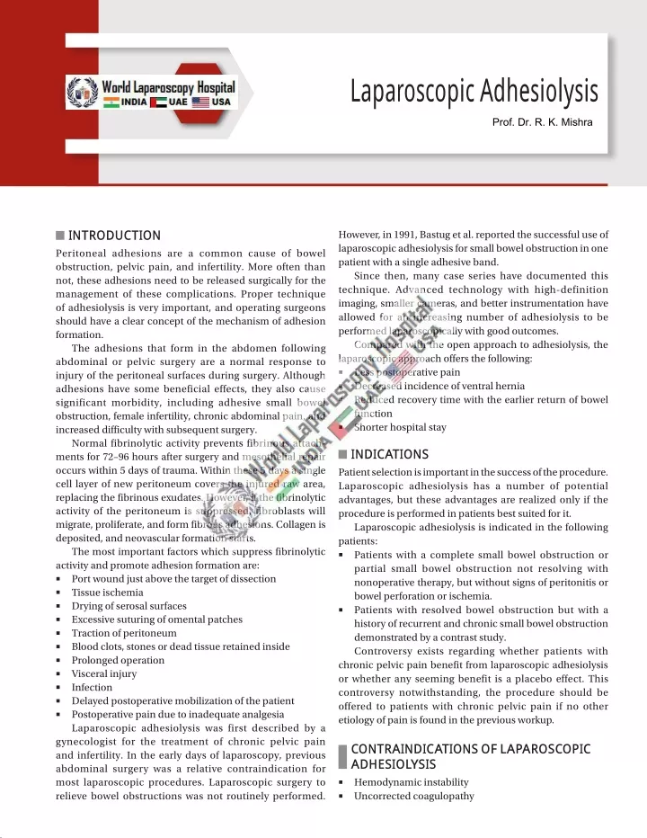
Peritoneal adhesions are a common cause of bowel obstruction, pelvic pain, and infertility. More often than not, these adhesions need to be released surgically for the management of these complications.

E N D
Laparoscopic Adhesiolysis Prof. Dr. R. K. Mishra INTRODUCTION Peritoneal adhesions are a common cause of bowel obstruction, pelvic pain, and infertility. More often than not, these adhesions need to be released surgically for the management of these complications. Proper technique of adhesiolysis is very important, and operating surgeons should have a clear concept of the mechanism of adhesion formation. The adhesions that form in the abdomen following abdominal or pelvic surgery are a normal response to injury of the peritoneal surfaces during surgery. Although adhesions have some beneficial effects, they also cause significant morbidity, including adhesive small bowel obstruction, female infertility, chronic abdominal pain, and increased difficulty with subsequent surgery. Normal fibrinolytic activity prevents fibrinous attach ments for 72–96 hours after surgery and mesothelial repair occurs within 5 days of trauma. Within these 5 days a single cell layer of new peritoneum covers the injured raw area, replacing the fibrinous exudates. However, if the fibrinolytic activity of the peritoneum is suppressed, fibroblasts will migrate, proliferate, and form fibrous adhesions. Collagen is deposited, and neovascular formation starts. The most important factors which suppress fibrinolytic activity and promote adhesion formation are: ■ Port wound just above the target of dissection ■ Tissue ischemia ■ Drying of serosal surfaces ■ Excessive suturing of omental patches ■ Traction of peritoneum ■ Blood clots, stones or dead tissue retained inside ■ Prolonged operation ■ Visceral injury ■ Infection ■ Delayed postoperative mobilization of the patient ■ Postoperative pain due to inadequate analgesia Laparoscopic adhesiolysis was first described by a gynecologist for the treatment of chronic pelvic pain and infertility. In the early days of laparoscopy, previous abdominal surgery was a relative contraindication for most laparoscopic procedures. Laparoscopic surgery to relieve bowel obstructions was not routinely performed. However, in 1991, Bastug et al. reported the successful use of laparoscopic adhesiolysis for small bowel obstruction in one patient with a single adhesive band. Since then, many case series have documented this technique. Advanced technology with highdefinition imaging, smaller cameras, and better instrumentation have allowed for an increasing number of adhesiolysis to be performed laparoscopically with good outcomes. Compared with the open approach to adhesiolysis, the laparoscopic approach offers the following: ■ Less postoperative pain ■ Decreased incidence of ventral hernia ■ Reduced recovery time with the earlier return of bowel function ■ Shorter hospital stay INDICATIONS Patient selection is important in the success of the procedure. Laparoscopic adhesiolysis has a number of potential advantages, but these advantages are realized only if the procedure is performed in patients best suited for it. Laparoscopic adhesiolysis is indicated in the following patients: ■ Patients with a complete small bowel obstruction or partial small bowel obstruction not resolving with nonoperative therapy, but without signs of peritonitis or bowel perforation or ischemia. ■ Patients with resolved bowel obstruction but with a history of recurrent and chronic small bowel obstruction demonstrated by a contrast study. Controversy exists regarding whether patients with chronic pelvic pain benefit from laparoscopic adhesiolysis or whether any seeming benefit is a placebo effect. This controversy notwithstanding, the procedure should be offered to patients with chronic pelvic pain if no other etiology of pain is found in the previous workup. CONTRAINDICATIONS OF LAPAROSCOPIC ADHESIOLYSIS Hemodynamic instability Uncorrected coagulopathy ■ ■
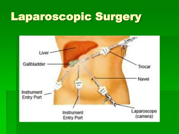
377 CHAPTER27: Laparoscopic Adhesiolysis ■ ■ ■ ■ Severe cardiopulmonary disease Abdominal wall infection Multiple previous upper abdominal procedures Late pregnancy iliac fossa should be introduced according to the baseball diamond concept after visualizing the target of dissection. Port should be in a position to provide an elevation angle of 30° and a manipulation angle of 60°, which is a standard and considered ergonomically better. Some gynecologists prefer suprapubic port, but with a suprapubic port, the elevation angle of the instrument and tubal structure is 90°, and hence lifting of ovary and tube may be difficult without grasping it. A three or four ports approach should be used if there is any difficulty in manipulation with two ports, especially in case of extensive adhesions (Figs. 1A and B). For adhesiolysis in right lower quadrant, the preferred port positions should be: ■ 10 mm umbilical (optical) ■ 5 mm suprapubic ■ 5 mm right hypochondrium A 30° telescope is employed in most instances, as this facilitates easier inspection of the deeper peritoneal cavity and abdominal organs. The secondary ports are inserted under laparoscopic vision. The selected site on the abdominal wall is identified by finger indentation of the parietal peritoneum. The open technique for trocar insertion is recommended if extensive adhesion is suspected. At the time of laparoscopic adhesiolysis, surgeon should try to be very gentle with the tubal structure and bowel so that readhesions are prevented and stricture of tube may not occur. Viewing of lateral pelvic organs is helped by the manipulation of mobile structures with a second port introduced through the left iliac fossa. Patient Position The anesthetized patient is placed on the operating table with the legs straight or in a lithotomy position if female. The lithotomy position will allow the gynecologists and assistant to work simultaneously, and uterine manipulation would be possible in case it is required. The thighs must not be flexed onto the abdominal wall as they would be in the full lithotomy position used for other open surgical, gynecological procedures. The operating table is tilted head up or down by approximately 15° depending on the main area of examination. Compression bandage may be used on legs during the operation to prevent thromboembolism, especially if the patient is in the lithotomy position. Position of the Surgical Team Before starting diagnostic laparoscopy, the best guess is made about the quadrant in which adhesions are more likely to be found. The surgeon should stand opposite to this quadrant to allow a direct view into this quadrant. If the pathology is more likely in the pelvic cavity the surgeon stands on left side of the patient. The first assistant, whose main task is to position the video camera, is also on the patient’s left side. The instrument trolley is placed on the patient’s left, allowing the scrub nurse to assist with placing the appropriate instruments in the operating ports. Television monitors are positioned on either side of the bottom end of the operating table at a suitable height for surgeon, anesthetists, as well as assistant to see the procedure. For adhesions involving other quadrants of the abdomen, the surgeon, assistants, and monitors are placed accordingly. PATHOGENESIS At the molecular level, adhesion formation involves a complex interaction of cytokines, growth factors, cell adhesion molecules, neuropeptides, and numerous other factors secreted by cells in or near the area of trauma. The early balance between fibrin deposition and degradation (i.e., fibrinolysis) appears to be a critical factor in the pathogenesis of adhesions. Port Position For the adhesiolysis of gynecological purposes, generally, one optical port in umbilicus and two 5 mm port in left and right A B Figs. 1A and B: Ports for pelvic adhesiolysis.
378 SECTION2: Laparoscopic General Surgical Procedures LAPAROSCOPIC ADHESIOLYSIS TECHNIQUE Animal studies have proved that laparoscopy leads to less adhesion formation compared to open surgery. The low adhesion forming tendency after laparoscopic surgery is because very less retraction is used, packing of the abdominal cavity is not required that can damage peritoneum. In laparoscopic surgery, there are fewer chances of drying of tissues because the inside environment is cutoff from outside. Also, the excellent visualization and magnification result in less likelihood of tissue injury and adhesions. In laparoscopy, port wound and wound at the target of dissection is far away from each other, so the chances of adhesions are less likely to the peritoneum because, for adhesions to form, both the raw layers which tends to adhere should be in contact. At least three ports should be used to perform laparoscopic adhesiolysis. After access and introduction of telescope, two other ports should be introduced according to the baseball diamond concept, keeping in mind the center of adhesions as a target of dissection. If the adhesions are thin and avascular, it is easily lysed, and the chances of recurrence are not much. In contrast, if adhesions are thick and highly vascular, then it is difficult to separate. These adhesions usually require the use of energy sources (ultrasonic dissector, unipolar, or bipolar). Regular achieving of hemostasis and meticulous sharp dissection with scissors is necessary (Figs. 2A and B). Adhesiolysis can be safely performed if dissection is done carefully through avascular planes. The laparoscopic approach precludes feeling through these adhesions. Accordingly, a general rule that can be followed in this setting is, if you can see through it, you can cut it. An atraumatic grasper is introduced to hold the adhesions or involved organs. It should be stretched gently, and boundaries of adhesion are identified. The avascular area is chosen with the closeup magnified view of the telescope. The opposite trocar on the side of the surgeon is used for scissors and adhesions should be cut close to the affected organ. Vascular adhesions should be coagulated using an electrosurgical instrument, preferably, bipolar. Scissors should be used only if flimsy avascular adhesions are found (Figs. 3A and B). Thick vascular adhesions should be first tried with blunt dissection, or otherwise must be coagulated before being cut. A suction irrigation instrument is good if blunt dissection is the main line of action. Try to get a sense of the tissue. Some patients have tissue that will tear easily, whereas others have tissue that readily A B Figs. 2A and B: Sharp dissection with scissors for bowel is involved. A B Figs. 3A and B: Sharp dissection with scissors if the bowel is involved.
379 CHAPTER27: Laparoscopic Adhesiolysis A B Figs. 4A and B: Tubo-ovarian mass with bowel involvement. ■ ■ permits blunt dissection. An individualized approach to each patient’s tissues is important. Bowel injury is not very uncommon during enterolysis and patients who have a history of the previous laparotomy should undergo a preoperative bowel preparation (Figs. 4A and B). If an inadvertent injury occurs, then enterorrhaphy can be accomplished with one layer closure using Vicryl. After adhesiolysis, some fluid can be left inside to which may help prevent a recurrence. Steroids and antihistamines have been tried, but are now used infrequently because of the adverse effect of delayed wound healing and high risk of dehiscence. Highmolecular weight dextran has also been tested to prevent readhesions because it is absorbed over a period of 7–10 days. Its osmotic effect draws the fluid into the peritoneal cavity, and so the mobile peritoneal organ floats, reducing adherence between intraperitoneal organs. Although the study in animals has demonstrated reduced postoperative adhesion, its efficacy is not fully confirmed. Adhesion barrier membranes have also been tried. These absorbable membranes separate the peritoneal lining from potentially likely to adhere to organs and thus prevent fibrous bands from different binding structures. Two such materials are Interceed and GoreTex. Interceed is an absorbable fabric of oxidized regenerated cellulose, and GoreTex is a nonabsorbable and nonreactive surgical membrane. Animal studies have demonstrated good results using these membranes. Inhibit the fibroblastic response to the tissue injury Involve recombinant tissue plasminogen activator and novel fibrinolytic Methods for preventing adhesions can be classified broadly as technical measures; physical barriers, which may be solid or liquid; and pharmacologic therapies. Gentle Tissue Handling A good surgical technique is the first defense against adhesion formation. Meticulous hemostasis and gentle, minimal tissue handling are important for limiting the extent of the initial peritoneal injury. Damage to the serosa can be prevented by minimizing trauma, bleeding, and ischemia and by keeping the surgical field moist with frequent irrigation to prevent tissues from drying out. Laparoscopy offers certain advantages over open abdominal surgery concerning adhesion formation. The abdominal incisions are small and there is less handling of tissue and exposure to foreign bodies, all of which may help to decrease tissue trauma, compared with laparotomy, and thus to reduce the risk for adhesion formation, especially to the abdominal wall. Physical Barriers Physical barriers include solid materials (absorbable sheets and nonabsorbable prosthetic materials) and viscous fluids introduced into the abdomen. All are aimed at keeping damaged peritoneal surfaces separated during the first 5–7 days after surgery until after reepithelialization has occurred. Although barriers do appear to limit the extent of adhesion formation, whether they improve clinically important outcomes by reducing the risks for intestinal obstruction, infertility, and chronic abdominal or pelvic pain is less. MEASURES FOR PREVENTING PERITONEAL ADHESIONS Fundamentals Methods for preventing adhesions are directed at the mechanisms of adhesion formation. ■ Minimize injury ■ Introduce a barrier between injured surfaces ■ Prevent coagulation of the serous exudate ■ Remove or dissolve the deposited fibrin Solid Barriers (Sheets) Two absorbable membrane sheets are commercially available. One is a sodium hyaluronatebased carbo xymethylcellulose sheet (Seprafilm) (Figs. 5A and B),
380 SECTION2: Laparoscopic General Surgical Procedures A B Figs. 5A and B: Seprafilm an adhesive barrier. A B Figs. 6A and B: Interceed an adhesive barrier. Liquid Barriers (Instillates) Polyethylene Glycol Polyethylene glycol (PEG) adhesion barrier (SprayGel and SprayShield) is a synthetic hydrogel that forms within seconds after simultaneous spray of two solutions of PEG based liquids onto targeted tissue. Crosslinking between the solutions forms an absorbable, flexible, and adherent gel barrier that remains intact for 5–7 days before degrading into its components, which are then resorbed and excreted through the kidneys. SprayGel is available in Europe but is not yet approved for use in the United States. and the second is an oxidized regenerated cellulose sheet (Interceed) (Figs. 6A and B). Both appear safe and effective for preventing adhesions between surfaces to which they are applied but are somewhat tricky to handle and do not avert adhesion formation at other sites within the abdomen. In addition, there is one nonabsorbable solid barrier [expanded polytetrafluoroethylene (ePTFE)] that has been found to prevent adhesions in clinical studies. Expanded Polytetrafluoroethylene Expanded polytetrafluoroethylene is a nonabsorbable, flexible prosthetic material used for a variety of surgical reconstructions. The ePTFE is trimmed to overlap the denuded area by 1 cm and sutured into place with nonabsorbable sutures, usually a 70 or 80 nylon or polypropylene. A small trial of patients having open myomectomy randomly assigned 28 subjects to the application of ePTFE suture over the uterine incision or to no barrier. At secondlook laparoscopy, more patients receiving ePTFE were adhesion free compared with untreated controls (55% vs. 7%). Icodextrin Solution A 4% isosmolar solution of icodextrin (Adept) is an alpha1,4 glucose polymer with the prolonged peritoneal residence. It is used as an irrigant during surgery (minimum 100 mL/30 min) and is the most promising intraoperative instillate for adhesion prevention. It can be used following laparoscopic adhesiolysis and is the only agent approved in the United States for preventing peritoneal adhesions in gynecologic laparoscopy.
381 CHAPTER27: Laparoscopic Adhesiolysis Hyaluronic Acid Solution, Gel, and Powder Hyaluronic acid is an anionic, nonsulfated glycosa minoglycan distributed in connective tissue. A systematic review identified four trials comparing hyaluronic acid containing solutions with placebo and found no difference in overall mean adhesions scores, but also found that the solutions significantly reduced the proportion of women with adhesions or the extent of adhesions at time of second look laparoscopy. formation after reproductive pelvic surgery. Fertil Steril. 1987;47(5):8646. 3. Easter DW, Cushieri A, Nathanson LK, LavelleJones M. The utility of diagnostic laparoscopy for abdominal disorders. Audit of 120 patients. Arch Surg. 1992;127(4):37983. 4. Eypasch E, Spangenberger W, Williams JI, Ure B, Neugebauer W, WoodDauphinee S, et al. Frfihe postoperative Verbesserung der Lebensqualitfit nach laparoskopischer Cholecystektomie. In: Hfiring R (Ed). Diagnostik und Therapie des Gallensteinleidens im Wandel der Zeit. Berlin: Blackwell; 1992. pp. 48191. 5. Fervers C. Die Laparoskopie mit dem Cystoskop. Ein Beitrag zur Vereinfachung der Technik und zur endoskopischen Strangdurchtrennung in der Bauchhohle. Med Klin Chir. 1933;178:288. 6. Fuchs KH, Freys SM, Heimbucher J, Thiede A. Laparoskopische Cholecystektomie—lohnt sich die laparoskopische Technik in “schwierigen” Ffillen? Chirurg. 1992;63:296304. 7. Klaiber C, Metzger A. Manual der laparoskopischen Chirurgie. Bern: Verlag Hans Huber; 1992. pp. 185206. 8. Kolmorgen K, Schulz AM. Results of laparoscopic lysis of adhesions in patients with chronic pelvic pain. Zentralbl Gynakol. 1991;113(6):2915. 9. Kresch AJ, Seifer DB, Sachs LB, Banner I. Laparoscopy in 100 women with chronic pelvic pain. Obstet Gynecol. 1984;64(5):6724. 10. Luciano AA, Maier DB, Koch EI, Nulsen JC, Whitman GF. A comparative study of postoperative adhesions following laser surgery by laparoscopy versus laparotomy in the rabbit model. Obstet Gynecol. 1989;74(2):2204. 11. Mecke H, Semm K, LehmannWillenbrock E. Pelviscopic adhesiolysis. Successes in the treatment of chronic abdominal pain caused by adhesions in the lower and middle abdomen. Geburtshilfe Frauenheilkd. 1988;48(3):559. 12. Meiser G, Waclawiczek HW, Heinerman M, Boeckl O. Der intermittierende inkomplette Dtinndarmileus—sono graphische Diagnostik und Trendbeobachtung. Chirurg. 1990;61:6516. 13. Nezhat CR, Nezhat FR, Metzger DA, Luicano A. Adhesion reformation after reproductive surgery by videolaseroscopy. Fertil Steril. 1990;53(6):100811. 14. Peters AA, TrimbosKemper GC, Admiraal C, Trimbos JB, Hermans J. A randomized clinical trial on the benefit of adhesiolysis in patients with intraperitoneal adhesions and chronic pelvic pain. Br J Obstet Gynaecol. 1992;99(1):5962. 15. Raf LE. Causes of abdominal adhesions in cases of intestinal obstruction. Acta Chir Scand. 1969;135:736. 16. Rapkin AJ. Adhesions and pelvic pain: a retrospective study. Obstet Gynecol. 1986;68(1):135. 17. Riedel HH, Haag GM. Late sequelae of appendectomy with special reference to adhesions in the lower abdomen, chronic abdominal pain and sterility. Zentralbl Gynakol. 1989;111(16):110112. 18. Riedel HH, LehmannWillenbrock E, Mecke H, Serum K. The frequency distribution of various pelviscopic (laparoscopic) operations, including complications rates—statistics of the Federal Republic of Germany in the years 1983–1985. Zentralbl Gynakol. 1989;111:7891. 19. Serum K. Operationslehre ftir endoskopische Abdomin alchirurgie. Stuttgart: Schattauer; 1984. 20. Serum K. Sichtkontrollierte Peritoneumperforation zur operativen Pelviskopie. Geburtshilfe Frauenheilkd. 1988;48: 4369. 21. Tavmergen EN, Mecke H, Serum K. Hfiufigkeit intraabdomineller Adhfisionen nach Pelviskopie und Laparotomie. Zentralb Gynakol. 1990;112:11639. 22. Troidl H, Spangenberger W, Dietrich A, Neugebauer F. Laparoskopische Cholecystektomie. Erste Erfahrungen und Ergebnisse bei 300 Operationen: eine prospektive Beobachtungsstudie. Chirurg. 1991;62:25765. Promotion of Bowel Motility As an adjunct to the physical separation of healing surfaces, maintenance of relative motion between healing surfaces, mimicking normal peristalsis and mobility, has been proposed to prevent or limit adhesion formation. In humans, impaired postoperative intestinal motility may cause a postoperative ileus, further exacerbated by postoperative pain management with opiates. The small intestine has impaired motility for 1–2 days postoperatively, whereas the colon has impaired motility for 2–3 days on average. A number of other agents have been applied in attempts to prevent postoperative peritoneal adhesions but are considered generally ineffective and possibly harmful. Crystalloid solutions should not be expected to prevent adhesion formation because of their short intraperitoneal time of residence, and this prediction is consistent with clinical observations installation of nonbarrier fluid into the peritoneal cavity after surgery is associated with numerous theoretical and practical concerns. Fluid overload may potentially lead to pulmonary edema and may cause abdominal pain and dyspnea. Fluid may potentially leak through laparoscopic incisions, which may cause distress to the patient and require frequent bandage replacement. Extravasation to the vulvar region has been reported in up to 2% of patients receiving dextran 70. Excess intraperitoneal fluid also may reduce opsonization of foreign cells, impair host cell phagocytosis, and lead to infectious complications. In one animal study, dextran significantly reduced adhesion formation but resulted in peritonitis rather than in abscesses as was observed with the instillation of saline. Other solutions instilled into the peritoneum to reduce adhesion formation have been evaluated and are ineffective. A systematic review found no significant benefit from the use of intraoperative irrigation or infusion of various drugs and liquids, including intraperitoneal steroids, dextran, or heparin. Antibiotic solutions also are ineffective for preventing adhesions, and, in rats, irrigation of the abdominal cavity with cefazolin and tetracycline resulted in increased formation of peritoneal adhesions. BIBLIOGRAPHY 1. De Wilde RL. Goodbye to late bowel obstruction after appendicectomy. Lancet. 1991;338:1012. 2. Diamond MP, Daniell JF, Feste J, Surrey MW, McLaughlin DS, Friedman S, et al. Adhesion reformation and de novo adhesion