Download
1 / 21
210 likes | 245 Views
Laparoscopic surgery has undergone rapid development in recent years. Laparoscopic cholecystectomy was first performed in 1985. Since the introduction of laparoscopic cholecystectomy into general practice in 1990, it has rapidly become the dominant procedure for gallbladder surgery.

E N D
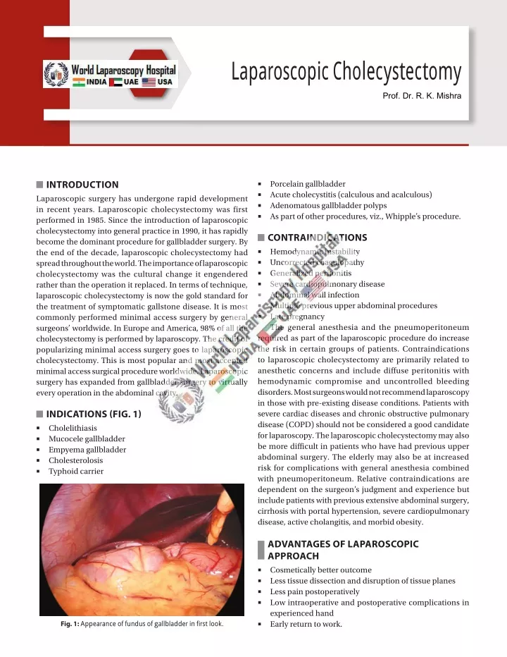
Laparoscopic C holecystectom y Prof. Dr. R. K. Mishra INTRODUCTION Laparoscopic surgery has undergone rapid development in recent years. Laparoscopic cholecystectomy was first performed in 1985. Since the introduction of laparoscopic cholecystectomy into general practice in 1990, it has rapidly become the dominant procedure for gallbladder surgery. By the end of the decade, laparoscopic cholecystectomy had spread throughout the world. The importance of laparoscopic cholecystectomy was the cultural change it engendered rather than the operation it replaced. In terms of technique, laparoscopic cholecystectomy is now the gold standard for the treatment of symptomatic gallstone disease. It is most commonly performed minimal access surgery by general surgeons’ worldwide. In Europe and America, 98% of all the cholecystectomy is performed by laparoscopy. The credit of popularizing minimal access surgery goes to laparoscopic cholecystectomy. This is most popular and most accepted minimal access surgical procedure worldwide. Laparoscopic surgery has expanded from gallbladder surgery to virtually every operation in the abdominal cavity. ■ ■ ■ ■ Porcelain gallbladder Acute cholecystitis (calculous and acalculous) Adenomatous gallbladder polyps As part of other procedures, viz., Whipple’s procedure. CONTRAINDICATIONS ■ Hemodynamic instability ■ Uncorrected coagulopathy ■ Generalized peritonitis ■ Severe cardiopulmonary disease ■ Abdominal wall infection ■ Multiple previous upper abdominal procedures ■ Late pregnancy The general anesthesia and the pneumoperitoneum required as part of the laparoscopic procedure do increase the risk in certain groups of patients. Contraindications to laparoscopic cholecystectomy are primarily related to anesthetic concerns and include diffuse peritonitis with hemodynamic compromise and uncontrolled bleeding disorders. Most surgeons would not recommend laparoscopy in those with pre-existing disease conditions. Patients with severe cardiac diseases and chronic obstructive pulmonary disease (COPD) should not be considered a good candidate for laparoscopy. The laparoscopic cholecystectomy may also be more difficult in patients who have had previous upper abdominal surgery. The elderly may also be at increased risk for complications with general anesthesia combined with pneumoperitoneum. Relative contraindications are dependent on the surgeon’s judgment and experience but include patients with previous extensive abdominal surgery, cirrhosis with portal hypertension, severe cardiopulmonary disease, active cholangitis, and morbid obesity. INDICATIONS (FIG. 1) Cholelithiasis Mucocele gallbladder Empyema gallbladder Cholesterolosis Typhoid carrier ■ ■ ■ ■ ■ ADVANTAGES OF LAPAROSCOPIC APPROACH ■ Cosmetically better outcome ■ Less tissue dissection and disruption of tissue planes ■ Less pain postoperatively ■ Low intraoperative and postoperative complications in experienced hand ■ Early return to work. Fig. 1: Appearance of fundus of gallbladder in first look.
178 SECTION2: Laparoscopic General Surgical Procedures TIMING OF SURGERY The optimal timing of surgery will depend upon the patient’s overall medical condition and underlying diagnosis. ■ Acute cholecystitis: Patients with acute cholecystitis, who are surgical candidates, should undergo cholecystectomy as soon as they are fully resuscitated, and the most qualified surgeon is available. Early cholecystectomy has been associated with improved patient outcomes compared with interval cholecystectomy. ■ Gallstone pancreatitis: Patients with gallstone pancreatitis have a high risk of recurrence within 30 days of their attack and therefore should undergo cholecystectomy during their index hospitalization after their symptoms resolve. ■ Obstructive jaundice: A history of pancreatitis or jaundice raises the likelihood of common bile duct (CBD) stones. Patients with isolated obstructive jaundice with or without cholangitis should either have their CBD cleared with urgent endoscopic retrograde cholangiopancreatography (ERCP) preoperatively or with cholangiography and laparoscopic common duct exploration at the time of surgery. If ERCP is successful, the patient should electively be scheduled for cholecystectomy. ■ Medical comorbidities: Identification of bleeding diatheses and poor cardiopulmonary reserve will help to identify patients who may not tolerate pneumoperitoneum or will have higher likelihood for conversion to open operation. In patients with multiple comorbidities or acute medical problem, a trial of antibiotics with consideration for a percutaneous cholecystostomy tube with delayed laparoscopic cholecystectomy in 6–8 weeks may be preferable. For young, otherwise healthy patients with gallstones but no evidence of pericholecystic inflammation or bile duct dilation, additional preoperative laboratory testing is not routinely necessary, unless a new clinical event has occurred, such as significant pain, fever, or jaundice, or the physical examination suggests that an abnormality is present. An electrocardiogram or chest radiograph is obtained as appropriate. For patients with complicated biliary tract disease, abnormal tests (e.g., liver function tests, amylase, and lipase) should be repeated to serve as a baseline for postoperative comparison. Coagulation tests are not routinely needed but may be obtained, if there is a reason to believe an abnormality may be present. Imaging Ultrasonography (US) of the right upper quadrant establishes the diagnosis of gallstones or abnormalities of the gallbladder wall. The US may also demonstrate CBD dilatation, stones, or evidence of acute inflammation of the gallbladder. Nuclear cholescintigraphy may be useful in cases in which the diagnosis remains uncertain after US. Magnetic resonance cholangiopancreatography (MRCP) may be useful to evaluate the common duct in patients with mild elevations of their transaminases or mild CBD dilatation on US. Endoscopic Retrograde Cholangiopancreatography If a patient has a dilated CBD, CBD stones, or jaundice, consideration should be given to a preoperative ERCP with clearing of the stones followed by laparoscopic cholecystectomy. In the absence of frank jaundice or cholangitis, mild abnormalities of liver enzymes and/ or bile duct dilation may also be managed effectively with intraoperative evaluation of the CBD, rather than preoperative ERCP. PREOPERATIVE INVESTIGATIONS Apart from routine preoperative investigations, in fit patients, the only investigations needed are ultrasound examination. Although practiced in some centers, intravenous cholangio- graphy may not be confirmative and is attended with the risk of anaphylactic reactions. Patient Position Patient is operated in the supine position with a steep head-up and left tilt. This typical positioning of laparoscopic cholecystectomy should be achieved once the pneumoperitoneum has been established. The patient is then placed in reverse Trendelenburg’s position and rotated to the left to give maximal exposure to the right upper quadrant. Laboratory Testing Laboratory testing in patients being evaluated for acute cholecystitis typically includes a complete blood count, liver enzymes, amylase, and lipase. Elevations in the serum total bilirubin and alkaline phosphatase concentrations are notcommon in uncomplicated cholecystitis, since biliary obstruction is limited to the gallbladder; if present, they should raise concerns about complicating conditions such as cholangitis, choledocholithiasis, or Mirizzi’s syndrome. However, mild elevations have been reported even in the absence of these complications and may be due to the passage of small stones, sludge, or pus. Position of Surgical Team (Fig. 2) The surgeon stands on the left side of the patient with the scrub nurse-camera holder-assistant. One assistant stands right to the patient and should hold the fundus-grasping forceps.
179 CHAPTER13: Laparoscopic Cholecystectomy Tasks Analysis ■ Preparation of the patient ■ Creation of pneumoperitoneum ■ Insertion of ports ■ ■ ■ ■ ■ ■ ■ ■ ■ ■ Diagnostic laparoscopy Dissection of visceral peritoneum Dissection of Calot’s triangle Clipping and division of cystic duct and artery Dissection of gallbladder from liver bed Extraction of gallbladder and any spilled stone Irrigation and suction of operating field Final diagnostic laparoscopy Removal of the instrument with complete exit of CO2 Closure of wound. Port Location (Figs. 3A to C) Four ports are used—optical (10 mm), one 5 mm, one 10 mm operating, and one 5.0 mm assisting port. The optical port is at or near the umbilicus and routinely a 30° laparoscope is used. Some surgeons who have started laparoscopy earlier are more comfortable with 0° telescope. The laparoscope is inserted through a 10-mm umbilical port and the abdominal cavity is explored for any obvious abnormalities. The secondary ports are then placed under direct visualization with the laparoscope. The surgeon places a 10-mm trocar in the midline and left to the falciform ligament at the epigastrium. Two 5-mm ports, one subcostal trocar in the right upper quadrant and another 5 mm trocar, lower, near the right anterior axillary line, are placed. Fig. 2: Position of surgical team. B A C Figs. 3A to C: (A) Ideal port position for laparoscopic cholecystectomy; (B and C) Port position of cholecystectomy.
180 SECTION2: Laparoscopic General Surgical Procedures LAPAROSCOPIC ANATOMY (FIG. 4) Laparoscopic view of the right upper quadrant on first look will demonstrate primarily the subphrenic spaces, abdominal surface of diaphragm, and diaphragmatic surface of the liver. The fundus of the gallbladder can be seen popping from the inferior surface of liver. The falciform ligament is seen as a prominent dividing point between left subphrenic space and the right subphrenic space. As the gallbladder is elevated and retracted toward the diaphragm, adhesion to the omentum or duodenum and transverse colon is seen. Exposure of Gallbladder and Cystic Pedicle (Figs. 5A to D) A grasper is used through the right lower 5-mm trocar to grasp the gallbladder fundus and retract it up over the liver edge to expose the entire length of the gallbladder. If there are adhesions to the gallbladder, they will need to be taken down using blunt and sharp dissection. With the entire gallbladder visualized, a second grasper is inserted through the other right upper quadrant trocar to grasp the gallbladder infundibulum and retract it up and to the right to expose the triangle of Calot. Careful evaluation of the anatomy reveals whether it is partially intrahepatic, on a mesentery, or possesses a Phrygian cap or any other odd shape. Hartmann’s pouch should be identified and seen to be funnel down to continue as tubular structure, the cystic duct. It is important to identify Hartmann’s pouch clearly, because most of the laparoscopic surgeons clip and divide the cystic duct, high on its termination at Hartmann’s pouch, rather than attempting to trace the cystic duct’s junction with the CBD. Dissection of the junction of cystic duct with the CBD increases the chance of traction injury and bleeding from small vessels and lymphatic. Fig. 4: Topographic anatomy of gallbladder: (1) Gallbladder; (2) Cystic artery; (3) Mscagni lymph node; (4) Proper hepatic artery; (5) Abdominal aorta; (6) Portal vein; (7) Gastroduodenal artery. A B C D Figs. 5A to D: Proper traction for exposure of cystic pedicle.
181 CHAPTER13: Laparoscopic Cholecystectomy A B C D Figs. 6A to D: Clearing adhesions by hook. Cystic artery can be seen, if attention is given, as it runs along the surface of gallbladder. A lymph node may be seen anterior to the cystic artery. The cystic artery gives off a small artery that supplies the cystic duct. This tiny twig often avulsed and bleeds at the time of creating window between artery and duct. This bleeding stops when the cystic duct is clipped. Adhesiolysis (Figs. 6A to D) Any adhesion should be cleared from the gallbladder. The surgeon uses a dissector through the epigastric trocar to tear the peritoneal attachments from the infundibulum. The attachments are taken down from high on the gallbladder, beginning laterally in order to help avoid injury to the CBD. Sharp dissection may be carried out with the help of scissors attached with monopolar current. At the time of separating adhesion, surgeon should try to be as near as possible toward gallbladder. The cystic pedicle is a triangular fold of peritoneum containing the cystic duct and artery, the cystic node, and a variable amount of fat. It has a superior and an inferior leaf, which are continuous over the anterior edge formed by the cystic duct (Fig. 7). An important consideration is the frequent anomalies of the structures contained between the two leaves (15–20%). The normal configuration is for an anterior cystic duct with the cystic artery situated posterosuperiorly and aris ing from the right hepatic artery usually behind the CBD. Fig. 7: Pledget can be used for adhesiolysis. Dissection of Cystic Pedicle (Figs. 8A to D) The dissection of the cystic pedicle can be carried out with two-handed technique. The dissection should be started with anteromedial traction by left hand grasper placed on the anterior edge of Hartmann’s pouch. The anteromedial traction by left hand will expose the posterior peritoneum. The peritoneum of the posterior leaf of the cystic pedicle is divided superficially as far back as the liver. Posterior leaf is better to dissect before anterior leaf because it is relatively less vascular and the bleeding, if any, will not soil the anterior peritoneum; whereas if anterior peritoneum is tackled first, it may make the dissection area of posterior peritoneum filled
182 SECTION2: Laparoscopic General Surgical Procedures A B C D Figs. 8A to D: Dissection of cystic pedicle. Separation of Cystic Duct from Artery (Figs. 9A to D) with blood making dissection of this area difficult. Once the visceral peritoneum is dissected, a pledget mounted securely in a pledget holder is used for blunt dissection. Once the cystic duct is visualized, the dissector can be used to create a window in the triangle of Calot between the cystic duct and cystic artery. This window should be created high near the gallbladder-cystic duct junction to avoid injury to the common duct. The separation of the cystic duct anteriorly from the cystic artery behind can be performed by a Maryland’s grasper by gently opening the jaw of Maryland between the duct and artery. The opening of the jaw of Maryland dissector should be in the line of duct never at right angle to avoid injury of artery behind. Sufficient length of the cystic duct and artery on the gallbladder side should be mobilized so that three clips can be applied. The electrosurgical hook may be inserted into the window and hooked around the cystic duct. With an up and down movement, the hook is used to clear as much tissue as possible from the duct nearer to the cystic duct–gallbladder junction. Tissue that is not dissected free from the duct is retracted by the hook away from all structures and is divided using active cutting current. Depending on the length of the duct, it is usually not necessary to dissect it all the way down to its junction with the CBD. In a similar fashion, the hook can be used to isolate the cystic artery for a length that is adequate enough to clip it. Critical View of Safety Anterior and posterior dissection continues with alternating inferolateral and superomedial retraction of the neck until the gallbladder is dissected away from the liver, creating a “window” crossed by two structures—the cystic duct and artery. This is the “critical view of safety (CVS)” that should be achieved prior to clipping or dividing any tubular structures. There is no need to dissect down to the cystic duct–CBD junction unless the cystic duct is very short. The cystic artery should be dissected in a similar fashion. Calot’s node, or the cystic duct lymph node, is usually encountered adjacent and anterior to the artery and can be a useful landmark. The surgeon should be aware of certain anatomic variations in order to avoid misidentification of structures. A common anomaly is the right hepatic artery looping onto the infundibulum and being mistaken for the cystic artery. A short cystic duct is also seen quite frequently and could drain into the right hepatic duct, the common duct, or a low-lying aberrant right sectoral duct. In the presence of severe, acute, or chronic inflammation, or with a large stone in the neck of the gallbladder, the infundibulum may be “tethered” to the hepatic duct, which may lead the surgeon to misidentify the CBD for the cystic duct.
183 CHAPTER13: Laparoscopic Cholecystectomy A B C D Figs. 9A to D: Cystic duct is separated from artery by creating a window. Clipping and Division of Cystic Artery (Figs. 10A to D) The cystic artery is clipped and then divided by harmonic scalpel (Figs. 10I and J). Two clips are placed proximally on the cystic artery and one clip is applied distally. The artery is then grasped with a duckbill grasper on the gallbladder wall and then divided between second and third clip. the risk of CBD injury in case of difficult anatomy. If a cholangiogram is to be performed, the cystic duct is isolated and occluded with a clip, which is placed high on the duct at its junction with the gallbladder. This will avoid leakage of contents from the gallbladder when the duct is opened. The scissors are used to incise the duct. The opening in the cystic duct is made on the anterosuperior aspect. Correct alignment of the cystic duct and infusion of saline facilitates insertion of ureteric catheter to perform cholangiography. Insertion is difficult, if the opening in the cystic duct is made too close to the gallbladder. The dissector is used to spread the incision to adequately dilate it for introduction of the cholangiogram catheter. The catheter is introduced through one of the 5-mm port. It is secured by either inflating a balloon or placing a clip to hold it in place. The catheter is then flushed with saline to ensure appropriate placement. All instruments are removed and a dynamic cholangiogram with real-time fluoroscopy is performed. The contrast medium should be injected slowly during screening and the patient should be in a slight Trendelenburg’s position with the table rotated slightly to the right. It is essential that the entire biliary tract be outlined. When the cholangiogram is completed, the catheter is removed, and two clips are placed proximally on the duct. The duct is then divided. Surgeon should ligate or clip cystic duct when he is sure up to the point of absolute certainness. Clipping and Division of Cystic Duct (Figs. 10E to L) After isolating the cystic duct and artery, the clipper is introduced through the epigastric port and at least two clips are placed on the proximal side of the cystic duct. Care is taken not to place the clips too low because retraction can tent up the CBD or cause it to be obstructed. Another clip is placed on the gallbladder side of the cystic duct, leaving enough distance between the clips to divide it. In a similar fashion, clips are placed on the cystic artery, two proximally and one on the gallbladder side of the artery. The harmonic scalpel are then used through the epigastric port to divide the cystic duct and artery between the clips. Both the jaw of scissors should be under vision. Intraoperative Cholangiogram (Figs. 11A and B) In many institutions, routine intraoperative cholangiogram (IOC) is performed. Routine cholangiogram decreases
184 SECTION2: Laparoscopic General Surgical Procedures ■ The main advantages of intraoperative cholangiography during cholecystectomy are: ■ Detection of CBD stone ■ Reduction of the incidence of residual CBD stone Delineation of the biliary anatomical variations at risk for bile duct injury Intraoperative cholangiogram is a highly sensitive tool for detecting choledocholithiasis, with an overall accuracy of A B C D F E H G Figs. 10A to H
185 CHAPTER13: Laparoscopic Cholecystectomy I J K L Figs. 10I to L Figs. 10A to L: Cystic duct and artery are clipped and divided. A B Figs. 11A and B: Intraoperative cholangiogram. (CBD: common bile duct; CHD: common hepatic duct) 95%. Routine intraoperative cholangiography can diagnose unsuspected CBD stone in 1–14% (average 5%) of patients without indications for ductal exploration. With increased experience, successful laparoscopic IOC can be achieved in 90–99% of cases, a rate similar to that of IOC during open cholecystectomy. The failure of laparoscopic IOC is due to: ■ The narrowness of the cystic duct ■ Cystic duct rupture ■ Obstructive cystic valves ■ Impacted cystic stones ■ Dye extravasation from cystic duct perforation Intraoperative Ultrasonography If there is any concern for choledocholithiasis (elevated bilirubin and dilated duct) or uncertain anatomy, the CBD should be imaged intraoperatively. This should occur after the first clip is placed on the cystic duct. IOC
186 SECTION2: Laparoscopic General Surgical Procedures or laparoscopic ultrasound (LUS) may be performed. Many studies focused on the role of intraoperative US in laparoscopic cholecystectomy. Intraoperative US is technically demanding, but is as accurate as IOC (88–100%) for screening CBD stone. The advantages of intraoperative US over IOC are: ■ Speedy ■ Safer ■ Multiple time use ■ Economical The greater sensitivity of intraoperative US exami nation concerned mainly small stones or debris in the CBD. The disadvantages of intraoperative US is that it is impossible to provide extended views of the intrahepatic and extrahepatic biliary tree, difficult to show passage of contrast into the duodenum and to identify bile duct injuries. the left hand grasper. The electrosurgical hook or harmonic scalpel is used with cautery through the epigastric port to dissect the gallbladder from the bed of the liver. Using a grasper, the gallbladder is first retracted right to expose and dissect the medial side of the attachment. The gallbladder is then retracted to the left and the lateral side is dissected. Using this back-and-forth action, the hook is used to dissect the gallbladder off the bed from inferior to superior until it is 90% removed from the liver. Holding the remaining portion of the gallbladder attached to the liver, the dissection bed and the clipped structures are evaluated and any active bleeding is stopped using heal of the hook or spatula. Gallbladder should be separated from the liver through the areolar tissue plane binding the gallbladder to the Glisson’s capsule lining the liver bed (Fig. 13). The actual separation can be performed with scissors with electrosurgical attachment or electrosurgical hook knife. Pledget can be used to remove the gallbladder from liver bed once a good plane of dissection is found. Perforation of the gallbladder during its separation is a common complication, which is encountered in 15% of cases. One should be careful at the time of dissection and if there is spillage of stone, each stone should be removed from the peritoneal cavity to avoid abscess formation in future. Ligation of Cystic Duct Although the majority of surgeons opt for clipping the cystic duct, before dividing it, this technique, though quick, is intrinsically unsound, as internalization of the metal clip inside the CBD over the ensuing months is well documented. There is report of internalization of clip and subsequent stone formation after many years. The internalized clip becomes covered with calcium bilirubinate pigment. For this reason, to tie the cystic duct using a catgut Roeder external slip knot should be done. Extraction of Gallbladder (Figs. 14A to D) The gallbladder is now freed from the liver and placed on top of the liver. The patient is returned to the supine position and the area of dissection and the upper right quadrant are irrigated and suctioned until clear. The gallbladder is extracted through the 11-mm epigastric operating port with the help of gallbladder extractor. The specimen can also be retrieved through the subxiphoid (epigastric) port site. We generally prefer epigastric port for removal of uncomplicated gallbladder. We feel that the subxiphoid site may be better for specimen extraction, as the subxiphoid site has a lower likelihood of hernia formation. Many surgeons use umbilical port for withdrawal of gallbladder. The umbilical incision Dissection of Gallbladder from Liver Bed (Fig. 12) The assistant places continual cephalad traction on the gallbladder fundus while the surgeon alternates medial and lateral anteroposterior traction on the infundibulum with Fig. 13: Glisson’s capsule lining the liver bed. Fig. 12: Separation of gallbladder from gallbladder bed.
187 CHAPTER13: Laparoscopic Cholecystectomy A B C D Figs. 14A to D: Extraction of gallbladder. Fig. 15: Extraction of gallbladder by gentle pull from outside. Fig. 16: Extraction of gallbladder by hiding the neck in cannula. has no muscle layers and is easy to extend with minimal pain and cosmetic sequelae. If gallbladder is removed through the umbilical port, the laparoscope is placed through the epigastric port and the gallbladder is visualized on the dome of the liver. A large grasper with teeth is placed through the umbilical port and used to grasp the gallbladder along the edge of the clipped cystic duct stump. The gallbladder is then exteriorized through the umbilical incision where it is held into position with a clamp (Fig. 15). First, the neck of the gallbladder should be engaged in the cannula and then cannula will withdraw together with neck of gallbladder held within the jaw of gallbladder extractor (Fig. 16). Once the port with the neck of the gallbladder is out, the neck is grasped with the help of a blunt hemostat and it should be pulled out with screwing. If gallbladder is of small size, it will come without much difficulty, otherwise small incision should be given over the
188 SECTION2: Laparoscopic General Surgical Procedures Fig. 17: Suction of bile to extract gallbladder. Fig. 18: Stone should be taken out to reduce volume of gallbladder. A Fig. 19: Stone may be crushed to reduce volume of gallbladder. neck of the gallbladder and suction irrigation instrument should be used to suck all the bile to facilitate easy withdrawal (Figs. 17 and 18). Sometimes big stones will not allow easy passage of gallbladder and in these situations, ovum forceps should be inserted inside the lumen of gallbladder through the incision of its neck and all the stone should be crushed (Fig. 19). When ovum forceps is used to remove the big stones from the gallbladder, care should be taken that gallbladder should be held loose to have room for forceps, otherwise it will perforate and all the stone may spill out (Figs. 20A and B). Extraction inside a bag is recommended as a safeguard against stone loss and contamination of the exit wound (Figs. 21 and 22). B Figs. 20A and B: Once the gallbladder is empty, gentle pull from outside will facilitate its extraction. Gallbladder Perforation during Laparoscopic Cholecystectomy During the dissection of the gallbladder, the laparoscopic surgeon may enter the gallbladder inadvertently, causing spillage of bile and/or stones. The hole in the gallbladder can be closed by incorporating it within the grasping forceps or may be sutured closed to prevent further spillage. The bile should be suctioned and irrigated. Bile spillage, when not
189 CHAPTER13: Laparoscopic Cholecystectomy A B Fig. 21: The spilled stone and torn gallbladder can be taken out with the help of endobag. Figs. 22A and B: Extracted spilled stone and torn gallbladder. accompanied by stone spillage and promptly evacuated, has not been shown to increase postoperative infections. These instruments are either placed through special low- friction trocars of small diameters (2–4 mm). Although mini-laparoscopic cholecystectomy requires the same four incisions as standard cholecystectomy, three of the incisions are smaller and therefore less invasive to the patient. The remaining umbilical incision has a standard size of 5–10 mm, which permits the use of a standard laparoscope and specimen extraction. About 10% of attempted mini- laparoscopic cholecystectomies require conversion, most often to standard laparoscopic cholecystectomy rather than open surgery. Ending of the Operation The instrument and then ports are removed. Telescope should be removed leaving gas valve of umbilical port open to let out all the gas. At the time of removing umbilical port, telescope should be again inserted and umbilical port should be removed over the telescope to prevent any entrapment of omentum. The wound is then closed with suture. Vicryl should be used for rectus and unabsorbable intradermal or stapler for skin. A single suture is used to close the umbilicus and upper midline fascial opening. Many laparoscopic surgeons routinely leave this fascial defect without ill defect. Some surgeons like to inject local anesthetic agent over port site to avoid postoperative pain. Sterile dressing over the wound should be applied. Since the first laparoscopic cholecystectomy was performed, it has met with widespread acceptance as a standard procedure using four trocars. The fourth (lateral) trocar is used to grasp the fundus of the gallbladder so as to expose Calot’s triangle. It has been argued that the fourth trocar is not necessary in most cases. The three-port technique is as safe as the standard four-port. The main advantages of the three-port technique are that it causes less pain, is less expensive, and leaves fewer scars. The three- port cholecystectomy should be performed by experienced laparoscopic surgeon because left hand movement is very important in this surgery. Bimanual skill and correct interpretation of anatomy is a must before proceeding for this technique. Three-port cholecystectomy is possible by the experienced laparoscopic surgeons. Laparoscopic Cholecystectomy in Acute Cholecystitis Impaction of a stone in the cystic duct will result in bile stasis in the gallbladder. Acute cholecystitis results from inflammation of the mucosa of the gallbladder, which may secondarily become infected. This inflammation can then evolve to hydrops of the gallbladder and gallbladder abscess. Complications such as necrosis, perforation, phlegmon, or peritonitis can result, if surgical intervention is not done in timely manner. In certain cases, acute cholecystitis can be acalculous, especially in diabetic patients or in the immediate postoperative period following other surgical interventions. In the elective setting, a laparoscopic cholecystectomy has become the standard of care; the laparoscopic approach to acute cholecystitis remains controversial. In past publications, acute cholecystitis was considered as a relative or absolute contraindication to a laparoscopic approach. The laparoscopic approach to acute cholecystitis is preferred in practice by a large number of experienced teams. Nowadays, laparoscopic approach is considered safe, if the intervention is performed in a timely fashion. If laparoscopy is considered in acute attack, a conversion rate of 20% demonstrates that the procedure is only pursued when felt to be safe. Mini-laparoscopic Cholecystectomy Mini-laparoscopy uses instruments that are thinner than the standard instruments (<3 versus 5–10 mm).
190 SECTION2: Laparoscopic General Surgical Procedures Laparoscopic cholecystectomy can be done safely in the setting of acute or gangrenous cholecystitis and should be done as soon as possible to fall within the “golden period” within 72 hours when edema is the primary manifestation of the inflammation. If the patient presents in a delayed fashion (>72 hours from onset of symptoms), the surgery can be performed if there are no contraindications to an open procedure, as conversion is a significant risk. In the face of acute cholecystitis with multiple comorbidities, it may be preferable to treat with intravenous antibiotics and possibly percutaneous drainage, with subsequent laparoscopic cholecystectomy 6–8 weeks later. ■ Acute cholecystitis is distinguished from biliary colic by the presence of at least two of the following signs: Right upper quadrant pain lasting for more than 24 hours Fever more than 37°C The presence of a palpable distended tender gallbladder Elevation of the WBC count >11,000/mm3 Ultrasonographic findings demonstrating thick ening of the gallbladder wall > 4 mm Pericholecystic fluid collection in ultrasound The studies have concluded that the ideal time for the surgical intervention is within 48–72 hours of the appearance of symptoms. Laparoscopic cholecystectomy, when performed early, leads to reduction in: ■ Operative difficulty ■ Conversion rate ■ Operative time One of the major determining factors is the conversion rate, an indicator of the actual difficulty of the procedure. Numerous studies have demonstrated a significantly lower conversion rate when the laparoscopic cholecystectomy is performed within the first 72 hours of the onset of symptoms as opposed to a delayed procedure after this period. In case of an interval cholecystectomy following medical therapy, it must be performed within a period of 8–12 weeks. It is demonstrated that interval cholecystectomy is associated with a higher morbidity rate than in early intervention. should be ideally performed during the second trimester of pregnancy where the operative risks are considered to be the lowest. Laparoscopic cholecystectomy during pregnancy has been reported in many series of randomized controlled trials, with no specific complications related to the laparoscopic approach. When laparoscopic surgery is performed in pregnant patients, the trocars need to be placed higher in the abdomen due to the enlarged uterus. The CO2 insufflation should be kept at the lowest possible pressure while maintaining an adequate working space. If CBD imaging is necessary, LUS should be used in place of IOC to limit radiation exposure. Single Incision Laparoscopic Cholecystectomy Single incision laparoscopic (SILS) cholecystectomy uses one rather than multiple skin incisions for trocar placement. Through a single skin incision typically made periumbilically, multiple trocars are placed through one or more fascial punctures. SILS has been most widely used in gallbladder surgery and has been extensively studied in comparison with standard laparoscopic cholecystectomy. Overall, SILS offers little benefit over standard laparoscopic surgery. A 2017 meta-analysis of 37 trials comparing SILS with conventional laparoscopic cholecystectomy showed that SILS is associated with better cosmesis, body image, and wound satisfaction and less postoperative pain, but a slightly longer operative time and a higher conversion rate. Additionally, the risk of incisional hernia is four times higher after SILS than after conventional laparoscopic cholecystectomy. z z z z z z POSTOPERATIVE CARE Most of the patients can be discharged 6 hours after the surgery or on the next day. The patient who has urinary retention, prolonged nausea, more pain, or difficulty in walking can be discharged after bowel sound. Three doses broad-spectrum antibiotic should be administered. First dose infused 1 hour before surgery, second and third should be administered on consecutive days. Few patients may complain pain over tip of shoulder after laparoscopic surgery. This is due to irritation of diaphragm from CO2. Tramadol hydrochloride is advised in these patients. Most of the patients are able to resume their normal day-to-day activity within 48–72 hours after surgery. Generally, the patient can return to their sedentary work within a week. The patient should have no activity restriction unless the umbilical incision was particularly large. Then, limited heavy lifting for a few weeks is advisable. Most patients are able to return to work within 1 week. Patients follow-up in clinic 2–4 weeks after their operation. Laparoscopic Cholecystectomy during Pregnancy There is a strong association between pregnancy and gallstones. In the past, elective laparoscopic cholecystectomy for symptomatic gallstones was deferred as much as possible until after pregnancy. For patients with severe biliary colic, however, waiting until after pregnancy can lead to repeated hospitalizations and complications. Laparoscopic cholecystectomy is relatively contraindicated during pregnancy due to the potentially adverse side effects on the fetus. However, when complications arise, a surgical intervention becomes mandatory. This intervention Postcholecystectomy Syndrome Postcholecystectomy syndrome is a complex of hetero- geneous symptoms including persistent abdominal pain
191 CHAPTER13: Laparoscopic Cholecystectomy COMPLICATIONS OF LAPAROSCOPIC CHOLECYSTECTOMY Early Complications ■ Common bile duct injury ■ Bile leak ■ Injury to viscera ■ Hemorrhage ■ Retained stones and abscess formation. and dyspepsia that recur and persist after cholecystectomy. In a prospective cohort study, only 60% of patients reported complete relief of abdominal pain at 12 weeks after cholecystectomy for uncomplicated symptomatic cholelithiasis. Treatment for postcholecystectomy syndrome is tailored to the specific cause of the symptoms. Diagnosis of the underlying problem causing postcholecystectomy syndrome usually requires imaging to look for retained or recurrent stones or identify a bile duct leak, stricture, or transection. This can be accomplished in most cases with ultrasound and/or computed tomography (CT) scanning followed by direct cholangiography or MRCP. MRCP provides a noninvasive alternative to direct cholangiography for evaluation of the biliary tract. Late Complications ■ Biliary strictures ■ Cystic duct clip stones. Laparoscopic Cholecystectomy and CBD Injury Incidence of iatrogenic CBD injury is 0.12% and 0.55% during open and laparoscopic cholecystectomy, respectively. Robotic Cholecystectomy Robot-assisted cholecystectomy has also been performed. Compared with conventional laparoscopic cholecystectomy, robotic cholecystectomy has not been found to be more effective or safer than for benign gallbladder diseases but incurs longer operative time and higher cost. The use of robotic-assisted extended cholecystectomy for gallbladder cancer is currently being evaluated. Common Causes of CBD Injury ■ Misinterpretation of anatomy—70% ■ Technical errors ■ Risk factors ■ Surgeon operates on image rather than reality. Visual psychological studies have shown that laparoscopic surgeon works on snap interpretation by brain and success or disaster depends on whether snaps are right or wrong. Snap interpretation will be wrong, if there is eyeball degradation. Surgeon should not dissect the cystic duct, if there is lack of initial identification and memory of key structure to the point of absolute certainty (Figs. 23A and B). Most important technical error is hilar bleeding and frantic attempts are made to control bleeding by electrosurgery. This frantic attack can catch the CBD and will be injured. In case of bleeding, surgeon should first apply pressure and if it does not stop, he should take suction irrigation Cholecystectomy by Natural Orifice Transluminal Endoscopic Surgery Transgastric or transvaginal cholecystectomy has been performed with natural orifice transluminal endoscopic surgery (NOTES). Compared with conventional surgery, NOTES offers true scarless cholecystectomy, which has the potential of minimizing postoperative patient discomfort. However, as an emerging technology, NOTES still faces many technical challenges. Currently, the transgastric cholecystectomy is no longer performed; only a few international centers continue to offer transvaginal cholecystectomy. A B Figs. 23A and B: Testing of common bile duct.
192 SECTION2: Laparoscopic General Surgical Procedures ■ and atraumatic grasper to find out exact point of bleeding. Electrosurgery should be used only when bleeding point is identified. Conservative treatment and biliary drainage for 6 weeks by ERCP stent insertion or PTBD (percutaneous transhepatic biliary drainage), if endoscopic stent application is not possible After several weeks: Reconstructive surgery Roux-en-Y choledocoduodenostomy or hepato- jejunostomy All the variation of cystic duct and artery should be memorized to avoid inadvertent injury of CBD. The variation in cystic artery and hepatic duct is shown in Figures 24A to D. ■ Types of CBD Injury Bismuth classification: ■ Type 1—CBD stump >2 cm ■ Type 2—CBD stump <2 cm ■ Type 3—hilar, right, and left duct confluence intact ■ Type 4—hilar, separation of right and left ducts ■ Type 5—injury to aberrant right duct ± CBD injury If complication of CBD injury is recognized intra- operatively, reconstructive surgery should be performed same time if surgeon has sufficient experience. For high complete transaction, Roux-en-Y hepaticojejunostomy is preferred. For lower complete injuries, primary suture repair over T-tube is better but long end of T-tube must not be exteriorized from same site. For partial injuries, insertion of T-tube and Roux-en-Y serosal patch should be thought. ■ ■ How to avoid injury? ■ Try to memorize initial anatomy of Calot’s triangle ■ A large distended gallbladder should be aspirated and lifted rather than grasped ■ Anterolateral traction is better than fundus pull to avoid tenting of CBD ■ Avoid meticulous dissection by energized instrument ■ Better to do skeletonization through pledget ■ During detachment of gallbladder from liver bed, maintain plane of adipose tissue ■ Use suction irrigation frequently. Tenting of the CBD should always be kept in mind at the time of dissection to avoid injury. Strategy to Handle Complication Recognized Postoperatively ■ USG + ERCP + MRCP ■ Fluid + electrolyte + systemic antibiotic A B Figs. 24A and B
193 CHAPTER13: Laparoscopic Cholecystectomy C D Figs. 24C and D Figs. 24A to D: Variation in cystic artery and hepatic duct. LAPAROSCOPIC CHOLECYSTECTOMY WITH REAL-TIME NEAR-INFRARED FLUORESCENT CHOLANGIOGRAPHY Sometimes, there are certain mistakes that can happen during laparoscopic cholecystectomy procedure. The most common mistake is usually the misidentification of the extrahepatic bile duct anatomy, which can lead to bile duct injury. At World Laparoscopy Hospital, now we have started using indocyanine green (ICG) during laparoscopic cholecystectomy (Fig. 25). However, there is something that can be down about the mistake, or basically, the mistake can be avoided. In order to avoid the mistake, it requires the doctor to use a technique or tools that will enhance or promote the easier recognition of the biliary anatomy Fig. 25: Indocyanine green (ICG) used in laparoscopic cholecystectomy.
194 SECTION2: Laparoscopic General Surgical Procedures in the intraoperative procedure by use of near-infrared fluorescence cholangiography or imaging. According to various studies on the use of the near- infrared cholangiography in laparoscopic cholecystectomy, the report showed that there was positive and successful intraoperative identification of the extrahepatic bile ducts. This reduced the likelihood of causing an injury to the bile ducts. This is, however, unlike the conventional imaging; whose results are usually not highly successful. When the doctor can identify the relevant anatomical structure, he is able to perform the operation easily without making any mistakes that can be costly and fatal to the patient. However, given this advantage, the procedure is yet to be adopted across the globe due to several reasons. One of the reasons is that the procedure takes times, involves exposure to radiation, and requires additional practitioners. Additionally, the ability to clearly identify the relevant anatomical structure or interpret an IOC clearly to some extent depends on the expertise of the surgeon. Understanding Laparoscopic Cholecystectomy Laparoscopic cholecystectomy is one of the most performed procedures in various countries. The procedure itself is usually associated with bile duct injury due to a poor intraoperative identification of the extrahepatic bile ducts. However, that complication is usually rare and actually it is associated with an incidence of 0.3–0.7%. However, it should be noted that when the mistake happens and there happens to be a bile duct injury, it can lead to various complications such as lower quality of life, morbidity, and mortality that are clinically relevant, and extra costs. Often an injury to the bile duct leads to leakage of bile and abdominal sepsis, which can lead to the obstruction or blockage of bile duct with obstructive jaundice. This will eventually lead to orthotopic liver transplantation. This complication should be detected as early as possible; however, if it is detected when it is late or has really advanced, it can be difficult to manage the bile ducts injuries since they will have caused severe damage, which progresses to biliary peritonitis, multiorgan failure, sepsis, and eventually death. Therefore, you need to understand that this is a condition that can be fatal, if not detected and managed early enough. As earlier mentioned, the intraoperative misidentification of the bile ducts anatomy is usually the main cause of bile ducts injury. Fluorescence cholangiography can help to reduce the risk of the bile duct, by helping to improve the orientation of the anatomy during laparoscopic cholecystectomy (Fig. 26). Understanding Real-time Infrared Cholangiography On the other hand, real-time infrared fluorescent cholangiography required is often performed after the intravenous injection of ICG is viewed as a better and new technique for easier recognition of the biliary anatomy (Fig. 27). One is not exposed to radiation and the procedure does not require any additional manpower. This makes it easy and actually a real-time and a flexible technique to use during laparoscopic cholecystectomy. Since the procedure is able to provide real-time visual or identification of the bile duct relevant anatomy, the outcome of the surgery can be improved leading to almost 100% success rates in various patients. According to a clinical study, results showed that infrared cholangiography provided an improved and real- time identification of the extrahepatic bile ducts during the phase of CVS dissection. The identification was faster, in fact, up to 10 minutes of earlier identification of the cystic duct and the CBD was recorded. One of the best things about this procedure is that the liver is able to quickly and exclusively clear ICG that is often intravenously administered before the technique is put into action. Still, according to the clinical study, it was established that the real-time identification of the hepatic and cystic arteries was obtained after a repeated dose of intravenous injection of ICG. Fig. 26: Indocyanine green fluorescence cholangiography. Fig. 27: Recognition of the biliary anatomy.
195 CHAPTER13: Laparoscopic Cholecystectomy Indocyanine Green Indocyanine green is also known as the fluorescent dye. As earlier mentioned, the liquid is usually injected intravenously; it is actually the one that helps to light the path for better and real-time identification of the biliary anatomy during the laparoscopic cholecystectomy. The near-infrared fluorescent cholangiography is usually performed using, as its name goes, a fluorescent imaging system that is usually used together with a laparoscope. The system must have a lighting system that provides light for both infrared and xenon rays. To perform this cholangiography technique, the ICG must be administered at least 45 minutes before the procedure. This dye has been used for quite some time now, in fact, since 1956. Today, it is widely being used in almost every hospital for surgical and various clinical purposes. Indocyanine green is able to provide better visualization of the biliary anatomy when it is illuminated by the infrared light, which makes it manifest fluorescence. When this happens the practitioner is able to clearly and in real time identify all the relevant biliary anatomy, thus enabling one to perform laparoscopic cholecystectomy better without causing injury to the bile ducts. Therefore, ICG acts as an imaging agent, which is sterile, water-soluble, and has a peak spectral absorption averaging at 805 nm in blood plasma or simply the blood. Since it is exclusively eliminated by the liver, it does not undergo enterohepatic recirculation. It only stays in the bile for about 8 minutes after injection. The removal of ICG depends on several factors, including the blood flow of the liver, biliary excretion, and parenchymal cellular function. Sometimes, fluorescence may not be detected 45 minutes after the injection of ICG. Therefore, when this happens, the second dose of 2.5 mg of ICG can be administered intravenously also. A second injection of ICG can also be given if there is anything regarding perfusion that cannot be clearly understood during the surgery. Other Advantages of Real-time Near-infrared Fluorescent Cholangiography Near-infrared fluorescent cholangiography improves the ability to analyze and identify the relevant biliary anatomic structure in a quick manner; therefore, it can help to reduce the time used in performing the procedure. Quicker assessment of the extrahepatic biliary anatomy is what is needed during laparoscopic cholecystectomy. The technique is usually able to shorten the operative time due to the fact there is no need to bring on board additional equipment and manpower, especially the radiology personnel for an X-ray. The technique also serves as a good teaching tool for practitioners who are able to identify the relevant extrahepatic structures in almost every patient. This helps them become more experienced with the procedure, which makes it easier for them to perform future procedures. The technique does not require any X-ray, therefore, no one is exposed to radiation. The ICG that is used is usually safe and does not cause any adverse reactions in patients. It is exclusively removed by the liver even before it undergoes enterohepatic recirculation. It is also a cost- effective technique that can help the patients to save some of their money that would otherwise have been used, if the technique was not used. However, near-infrared fluorescent technique comes with some limitations. The common one is that it lacks the standardization of the fluorescence imaging systems. This is quite a drawback for this technique because standardization is needed for the determination of the objective assessments during any fluorescence-guided surgery like the laparoscopic cholecystectomy. The differences in fluorescence cholangio- graphy make it difficult for the comparisons between fluorescence cholangiography studies. CONCLUSION The real-time near-infrared fluorescence cholangiography is new and better technology that can help with the identification of the vital biliary anatomy. Several studies have been done on the technique and have been proven to be good for laparoscopic cholecystectomy. It helps to reduce the risk of the bile duct injury by proving clear vision of the vital extrahepatic structures. The reduced risk helps to avoid various complications that can arise from the injury of the bile duct, which sometimes can be fatal to a patient. Exploration of the Biliary Anatomy Practitioners always report their experience with real-time near infrared cholangiography using ICG. Most of the times when it comes to laparoscopic cholecystectomy, feasibility and safety of the technique are usually well reported. The reports are quite interesting when it comes to robot single-site cholecystectomy (RSSC) where the ICG is still administered before the surgery and the biliary anatomy can be analyzed before any dissection is made. This is quite different for cholangiography requiring extensive dissection of the cystic duct. Therefore, using ICG during RSSC can help to analyze the extrahepatic biliary anatomy better and this could help to reduce the bile duct injury due to the misinterpretation of the biliary anatomy. However, there are no data to prove this statement. BIBLIOGRAPHY 1. Airan M, Appel M, Berci G, Coburg AJ, Cohen M, Cuschieri A, et al. Retrospective and prospective multi-institutional laparoscopic cholecystectomy study by the Society of American Gastrointestinal Endoscopic Surgeons. Surg Endosc. 1992;6:169-76. 2. Barkum AN, Barkun JS, Fried GM, Ghitulescu G, Steinmetz O, Pham C, et al. Useful predictors of bile duct stones in patients undergoing laparoscopic cholecystectomy. Ann Surg. 1994; 220:32-9.
196 SECTION2: Laparoscopic General Surgical Procedures 3. Bartlett DL, Fong Y, Fortner JG, Brennan MF, Blumgart LH. Long- term results after resection for gallbladder cancer: implications for staging and management. Ann Surg. 1996;224:639-46. 4. Berci G, Morgenstern L. Laparoscopic management of common bile duct stones. Surg Endosc. 1994;8:1168-75. 5. Berrevoet E, Biglari M, Sinove Y, De Baardemaeker L, Troisi R, de Hemptinne B. Outpatient laparoscopic cholecystectomy in Belgium: what are we waiting for? Acta Chir Belg. 2006;106:537-40. 6. Branum G, Schmitt C, Baillie J, Suhoclri P, Baker M, Davidoff A, et al. Management of major biliary complications after laparoscopic cholecystectomy. Ann Surg. 1993;217:532-41. 7. Calik A, Topaloglu S, Topcu S, Turkyilmaz S, Kucuktulu U, Piskin B. Routine intraoperative aspiration of gallbladder during laparoscopic cholecystectomy. Surg Endosc. 2007;21(9):1578-81. 8. Callagham J. Twenty-five years of gallbladder surgery in a small rural hospital. Am J Surg. 1995;169:313-5. 9. Champault A, Vons C, Dagher I, Amerlinck S, Franco D. Low-cost laparoscopic cholecystectomy. Br J Surg. 2002;89:1602-7. 10. Chijiiwa K, Nakano K, Ueda J, Noshiro H, Nagai E, Yamaguchi K, et al. Surgical treatment of patients with T2 gallbladder carcinoma invading the subserosal layer. J Am Coll Surg. 2001;192:600-7. 11. Clavies PA, Sanabria JR, Strasberg SM. Proposed classification of complications of surgery with examples of utility in cholecystectomy. Surgery. 1992;111:518-6. 12. Cleary SP, Dawson LA, Knox JJ, Gallinger S. Cancer of the gallbladder and extrahepatic bile ducts. Curr Probl Surg. 2007;44:396-482. 13. Coburn NG, Cleary SP, Tan JC, Law CH. Surgery for gallbladder cancer: a population-based analysis. J Am Coll Surg. 2008; 207:371-82. 14. Cullen DJ, Apolone G, Greenfiled S, Guadagnoli E, Cleary P. ASA physical status and age predict morbidity after three surgical procedures. Ann Surg. 1994;220:3-9. 15. Davies MG, O’Broin E, Mannion C, McGinley J, Gupta S, Shine MF, et al. Audit of open cholecystectomy in a district general hospital. Br J Surg. 1992;79:314-6. 16. de Aretxabala XA, Roa IS, Burgos LA, Araya JC, Villaseca MA, Silva JA, et al. Curative resection in potentially resectable tumours of the gallbladder. Eur J Surg. 1997;163:419-26. 17. Deziel DJ, Millikan KW, Economou SG, Doolas A, Ko ST, Airan MC. Complications of laparoscopic cholecystectomy: a national survey of 4,292 hospitals and an analysis of 77,604 cases. Am J Surg. 1993;165:9-14. 18. Deziel DJ. Complications of cholecystectomy. Surg Clin N Am. 1994;74:809-23. 19. Dixon E, Vollmer CM Jr, Sahajpal A, Cattral M, Grant D, Doig C, et al. An aggressive surgical approach leads to improved survival in patients with gallbladder cancer: a 12-year study at a North American Center. Ann Surg. 2005;241:385-94. 20. Driessen PJHA, Pradhan GN. Laparoscopic cholecystectomy in a small rural hospital. CJRM. 2000;5:70-3. 21. Escarce JJ, Shea JA, Chen W, Qian Z, Schwartz JS. Outcomes of open cholecystectomy in the elderly: a longitudinal analysis of 21,000 cases in the prelaparoscopic era. Surgery. 1995;117:156-64. 22. Fong Y, Heffernan N, Blumgart LH. Gallbladder carcinoma discovered during laparoscopic cholecystectomy: aggressive reresection is beneficial. Cancer. 1998;83:423-7. 23. Fried GM, Clas D, Meakins JL. Minimally invasive surgery in the elderly patient. Surg Clin North Am. 1994;74:375-87. 24. Galandiuk S, Mahid SS, Polk Jr HC, Turina M, Rao M, Lewis JN. Differences and similarities between rural and urban operations. Surgery. 2006;140:589-96. 25. Ganey JB, Johnson PA Jr, Prillaman PE, McSwain GR. Cholecystectomy: clinical experience with a large series. Am J Surg. 1986;151:352-7. 26. Gilliland TM, Traverso LW. Modern standards for comparison of cholecystectomy with alternative treatments for symptomatic cholelithiasis with emphasis on long-term relief of symptoms. Surg Gynecol Obst. 1990;170:39-44. 27. Girometti R, Brondani G, Cereser L, Como G, Pin MD, Bazzocchi M, et al. Post-cholecystectomy syndrome: spectrum of biliary findings at magnetic resonance cholangiopancreatography. Br J Radiol. 2010;83:351. 28. Gupta A, Agarwal PN, Kant R, Malik V. Evaluation of fundus first laparoscopic cholecystectomy. JSLS. 2004;8:255-8. 29. Hainsworth PJ, Rhodes M, Gompertz RHK, Armstrong CP, Lennard TWJ. Imaging of the common bile duct in patients undergoing laparoscopic cholecystectomy. Gut. 1994;35:991-5. 30. Haynes JH, Guha SC, Taylor SG. Laparoscopic cholecystectomy in a rural family practice: the Vivian, LA, experience. J Fam Pract. 2004;53:205-8. 31. Herzog U, Messmer R, Sutter M, Tondeffi R. Surgical treatment for cholelithiasis. Surg Gynecol Obstet. 1992;175:238-42. 32. Jatzko GR, Lisborg PH, Pertl AM, Stettner HM. Multivariate comparison of complications after laparoscopic chole cystectomy and open cholecystectomy. Ann Surg. 1995;221:381-6. 33. Kondo S, Nimura Y, Hayakawa N, Kamiya J, Nagino M, Uesaka K. Extensive surgery for carcinoma of the gallbladder. Br J Surg. 2002;89:179-84. 34. Kumar TS, Saklani AP, Vinayagam R, Blackett RL. Spilled gallstones during laparoscopic cholecystectomy: a review of literature. Postgraduate Med J. 2004;80:77-9. 35. Lamberts MP, Den Oudsten BL, Gerritsen JJ, Roukema JA, Westert GP, Drenth JPH, et al. Prospective multicentre cohort study of patient-reported outcomes after cholecystectomy for uncomplicated symptomatic cholecystolithiasis. Br J Surg. 2015;102:1402-9. 36. Leeder PC, Matthews T, Krzeminska K, Dehn TC. Routine day- case laparoscopic cholecystectomy. Br J Surg. 2004;91:312-6. 37. Macintyre IM, Wilson RG. Laparoscopic cholecystectomy. Br J Surg. 1993;80:552-9. 38. Mayol J, Alvarez Fernandez-Represa J. Imaging of the common bile duct (Letter). Gut. 1994;35:1773. 39. Mayol J, Tamayo FJ, Ortiz-Oshiro E, Ortega Lopez D. Resultados de la colecistectomia laparoscopica en pacientes ancianos. Or Esp. 1995;55:45-7. 40. Mayol J, Vincent-Hamelin E, Sarmiento JM, Oshiro EO, Diaz- Gonzalez J, Tamayo FJ, et al. Pulmonary embolism following laparoscopic cholecystectomy: report of two cases and review of the literature. Surg Endosc. 1994;8:214-7. 41. McSherry CK, Glenn F. The incidence and causes of death following surgery for non-malignant biliary tract disease. Ann Surg. 1980;191:271-5. 42. McSherry CK. Laparoscopic management of common bile duct stone [Editorial]. Surg Endosc. 1994;8:1161-2. 43. Mrozowicz A, Polkowski W. Initial three years’ experience with laparoscopic cholecystectomy in a district hospital: evaluation of early results and operative measures. Ann Univ Mariae Curie Sklodowska [Med]. 2004;59:26-31. 44. Muhe E. Die erste Cholezystektomie durch das Laparoskop. Langeb Arch Klin Chir. 1986;369:804. 45. Nakamura S, Sakaguchi S, Suzuki S, Muro H. Aggressive surgery for carcinoma of the gallbladder. Surgery. 1989;106:467-73. 46. Nakeeb A, Tran KQ, Black MJ, Erickson BA, Ritch PS, Quebbeman EJ, et al. Improved survival in resected biliary malignancies. Surgery. 2002;132:555-63. 47. Nimura Y, Hayakawa N, Kamiya J, Maeda S, Kondo S, Yasui A, et al. Hepato pancreatoduodenectomy for advanced carcinoma of the biliary tract. Hepatogastroenterology. 1991;38:170-5.
197 CHAPTER13: Laparoscopic Cholecystectomy 48. Parkar RB, Thagana NG, Baraza R, Otieno D. Experience with laparoscopic surgery at the Aga Khan Hospital, Nairobi. East Afr Med J. 2003;80:44-50. 49. Patel SC, Bhatt JR. Laparoscopic cholecystectomy at the Aga Khan Hospital, Nairobi. East Afr Med J. 2000;77:194-8. 50. Reddick EJ, Olsen DO. Laparoscopic laser cholecystectomy. Surg Endosc. 1989;3:131-3. 51. Shirai Y, Yoshida K, Tsukada K, Muto T. Inapparent carcinoma of the gallbladder. An appraisal of a radical second operation after simple cholecystectomy. Ann Surg. 1992;215:326-31. 52. Smith N, Max MH. Gallbladder surgery in patients over 60: is there an increased risk? South Med J. 1987;80:472-4. 53. Southern Surgeons group. A prospective analysis of 1518 laparo- scopic cholecystectomies. N Engl J Med N. 1991;324:1073-8. 54. Tan JT, Suyapto DR, Neo EL, Leong PS. Prospective audit of laparoscopic cholecystectomy experience at a secondary referral centre in South Australia. ANZ J Surg. 2006;76:335-8. 55. Taylor OM, Sedman PC, Jones BM, Royston CM, Arulampalam T, Wellwood J. Laparoscopic cholecystectomy without operative cholangiogram: 2038 cases over 5-year period in two district general hospitals. Ann R Coll Surg Engl. 1997;79:376-80. 56. Todoroki T, Kawamoto T, Takahashi H, Takada Y, Koike N, Otsuka M, et al. Treatment of gallbladder cancer by radical resection. Br J Surg. 1999;86:622-7. 57. Vagenas K, Karamanakos SN, Spyropoulos C, Panagiotopoulos S, Karanikolas M, Stavropoulos M. Laparoscopic chole cystectomy: a report from a single center. World J Gastroenterol. 2006; 12:3887-90. 58. van Dijk AH, Wennmacker SZ, de Reuver PR, Latenstein CSS, Buyne O, Donkervoort SC, et al. Restrictive strategy versus usual care for cholecystectomy in patients with gallstones and abdominal pain (SECURE): a multicentre, randomised, parallel- arm, non-inferiority trial. Lancet. 2019;393:2322. 59. Voyles CR, Sanders DL, Hogan R. Common bile duct evaluation in the era of laparoscopic cholecystectomy. 1050 cases later. Ann Surg. 1994;219:744-52. 60. Wakai T, Shirai Y, Hatakeyama K. Radical second resection provides survival benefit for patients with T2 gallbladder carcinoma first discovered after laparoscopic cholecystectomy. World J Surg. 2002;26:867-71. 61. Wang SJ, Fuller CD, Kim JS, Sittig DF, Thomas CR Jr, Ravdin PM, et al. Prediction model for estimating the survival benefit of adjuvant radiotherapy for gallbladder cancer. J Clin Oncol. 2008;26:2112-7. 62. Wherry DC, Marohn MR, Malanoski MP, Hetz SP, Rich NM. An external audit of laparoscopic cholecystectomy in the steady state performed in medical treatment facilities of the Department of Defense. Ann Surg. 1996;224:145-54. 63. Williams Jr LF, Chapman WC, Bonau RA, McGee Jr EC, Boyd RW, Jacobs JK. Comparison of laparoscopic cholecystectomy with open cholecystectomy in a single center. Am J Surg. 1993;165:459-65. 64. Wittgen CM, Andrus JP, Andrus CH, Kaminski DL. Chole- cystectomy. Which procedure is best for the high-risk patient? Surg Endosc. 1993;7:395-9. 65. Yamaguchi K, Chijiiwa K, Shimizu S, Yokohata K, Tsuneyoshi M, Tanaka M. Anatomical limit of extended cholecystectomy for gallbladder carcinoma involving the neck of the gallbladder. Int Surg. 1998;83:21-3. 66. Yamashita Y, Kurohiji T, Kakegawa T. Evaluation of two training programs for laparoscopic cholecystectomy: incidence of major complications. World J Surg. 1994;18:279-85.