Download
1 / 1
10 likes | 78 Views
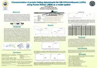
HD Domain. LNR Domain. A. B. C. 1:5. 2:4. 3:6. 5:1 Best. Post DTT incubation. LNRB_delBC. LNRB_int. LNRB_orig. Post DTT incubation. LNRB_delAB. LNRB_short. Characterization of protein folding determinants for LIN-12/Notch-Repeats (LNRs) using Human Notch1 LNR-B as a model system

E N D
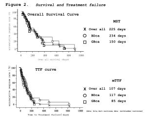
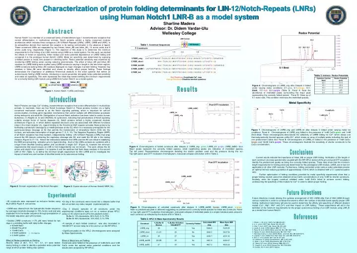
HD Domain LNR Domain A B C 1:5 2:4 3:6 5:1 Best Post DTT incubation LNRB_delBC LNRB_int LNRB_orig Post DTT incubation LNRB_delAB LNRB_short Characterization of protein folding determinants for LIN-12/Notch-Repeats (LNRs) using Human Notch1 LNR-B as a model system Sharline Madera Advisor: Dr. Didem Vardar-Ulu Wellesley College Abstract Redox Potential Human Notch1 is a member of a conserved family of heterodimeric type 1 transmembrane receptors that control differentiation in multicellular animals. Notch proteins exhibit a highly conserved modular architecture which includes three contiguous LIN-12/Notch-Repeats (LNRs), LNRA, LNRB and LNRC, in its extracellular domain that maintain the receptor in its resting conformation in the absence of ligand. These conserved LNRs are separated by two linkers, linker_AB and linker_BC, 10 amino acids and 5 amino acids long, respectively (Figure 1). The objective of this study is to determine the minimum requirements for the folding of an LNR module using LNRB as a model system. For this work, we studied the effects of metal ion specificity, linker residue and redox potential dependence on LNRB folding and compared it to the prototype human Notch1 LNRA. Metal ion specificity was determined by exposing unfolded protein to metal ions present in refolding buffer. Redox potential sensitivity was examined by monitoring LNRB folding under varying reducing environments. The effect of linker_AB and linker_BC residues on LNRB folding were studied using LNRB constructs varying in length in the two linker regions. LNRB constructs lacking linker_BC residues displayed no major changes in protein folding. However, key residues in linker_AB were identified and shown to directly affect proper folding. These findings demonstrate the importance of additional N-terminal residues to the initial cysteine that define an autonomously folding LNRB module, introducing a crucial parameter alongside redox potential sensitivity and metal ion specificity. This work represents the initial step toward defining the minimum requirements for a correctly folding LNR module using LNRB from human Notch1 as a model system. 15:1 Table 1. Construct Sequences 2:1 30:1 5:1 10:1 B Table 3. Intrinsic Redox Potentials Red Residues: Coordinate Ca2+ ions Orange Residues: Disulfide bonded cysteines linker_BC linker_AB CC CC CC SLNFNDPWKN QRAEGQ Figure 6. Chromatograms of LNRB_orig after Dialysis 3 folded under varying redox conditions: 2:1-grey, 5:1-orange, 10:1-black, 15:1-red, 30:1-purple (Table 3). Panel A: Note the abundance of misfolded peaks surrounding the major peak representing the correctly folded protein. Panel B: Close up of 5:1 redox ratio. This condition minimizes the misfolded species. LNRA Results LNRC LNRB Figure 1. Human Notch 1 LNRs and linkers. Introduction Metal Specificity Notch Proteins are large Ca2+ binding, transmembrane receptors that control differentiation in multicellular animals. In mammals, there are four Notch homologs: Notch1-4. These proteins function via a highly conserved mechanism referred to as the Notch signaling pathway, which is important for cell-cell communication, involving gene regulation mechanisms that control multiple cell differentiation processes during embryonic and adult life. Deregulation of normal Notch activation has been noted in certain human leukemias, (1) Alagille (2, 3) and CADASIL (4) syndromes, indicating that perturbations of Notch signaling underlie several forms of human diseases (5). Notch proteins exhibit a highly conserved modular architecture (Figure 2), in which distinct repeated structural units are associated with different functional roles in the intact receptor (6). Ligand binding to the N-terminal EGF-repeats activates these proteins by facilitating a proteolytic cleavage by a metalloprotease at site S2, which is a necessary prerequisite for the gamma-secretase cleavage at S3 that permits the translocation of intracellular Notch (ICN) into the nucleus, and activates transcription of target genes (7, 8, 9, 10). The Negative Regulatory Region (NRR) of all Notch receptors has three tandem, independently folding LIN-12/Notch Repeats (LNRs) that wrap around the HD domain containing the regulatory cleavage site S2, and mask the S2 site in the resting receptor (Figure 3) (11-13). Hence the interactions between the LNRs and the HD are critical in stabilizing the NRR and preventing activation prior to ligand binding. Each of the LNRs contains six cysteines with a unique three disulfide bonding pattern and coordinate a single Ca2+ (Figure 3), however the minimum requirements that would ensure an LNR to fold independently are not known. This work utilizes the 32 amino acid stretch from cysteine 1 to cysteine 6 and the residues that flank these residues in the second LNR of hN1 (Table 1), to define the minimum length requirement for hN1 LNRB and to investigate the impact of metal ions and number of disulfide bonds on its autonomous folding. B A CaCl2 10 mMCaCl2 ZnCl2 1 mMCaCl2 Figure 7. Chromatograms of LNRB_orig and LNRB_int after dialysis 3 folded under varying metal ion conditions. Panel A: Chromatograms of LNRB_orig folded in the presence of 1mM CaCl2-green and 1mM ZnCl2-blue. These chromatogramsdemonstrate the selectivity of LNRB folding for Ca2+ which shows one thermodynamically favored species unlike Zn2+ which shows an array of multiple peaks indicating the lack of one predominant native fold. Panel B: Chromatograms of LNRB_int folded in the presence of 1mM CaCl2-brown and 10mM CaCl2-purple. These chromatograms illustrate the sensitivity of shorter constructs to the CaCl2 concentration. Figure 4. Chromatograms of folded constructs after dialysis 3: LNRB_orig- green, LNRB_int- purple, LNRB_delBC- blue. Major peaks represent the correctly folded species, small neighboring peaks are indicative of misfolded species. Top left panel: Representative chromatogram detailing the elution gradient used and the pressure during the run. Top right panel: post DTT incubation chromatogram, note peak collapse and elution shift in Table 2. Conclusions Current results indicate the importance of linker_AB on proper LNR folding. Verification of the length of each construct via mass spectrometry coupled with the RP-HPLC elution shifts pre and post DTT incubation identify the predominant peaks as being the correctly folded species. These data show that the minimum length requirements for folding previously determined for the prototypical LNR module, LNRA, are in fact not applicable to all other LNR modules. Furthermore, folding conducted under various redox potentials provide an optimal intrinsic reducing potential of approximately -4.5mV, which is obtained with a 5:1 cysteine:cystine ratio. Further optimization of folding conditions provided by metal specificity experiments show that a proper folding module cannot be obtained at low CaCl2 concentrations of only 1mM for shorter constructs. Similarly, even the longest construct refolded under 1mM ZnCl2 failed to achieve correct folding, underscoring the specificity of this module for Ca2+ in order to obtain proper folding. Figure 2. Domain organization of the Notch Receptor. Figure 3. Crystal structure of Human Notch2 NRR (14). Future Directions Experimental Future directions include altering the cysteine arrangement of hN1 LNRB after that of hN4 LNRA through various mutations in order to correlate the extent to which the number of disulfide bonds specify proper LNR folding. Isothermal Calorimetry will also be used to determine the affinity and specificity of different divalent metals (Ca2+, Mg2+, Mn2+ and Zn2+) and their impact on LNR folding. These experiments will aid in the definition of the minimum requirements for the proper autonomous folding of an LNR module using LNR_B as a model from human Notch1. • All constructs were expressed as inclusion bodies using BL21(DE3) PlysS E. coli cell line. • LNRB was cleaved from the hydrophobic leader sequence by cyanogen bromide cleavage in 70% formic acid and was separated from the leader sequence through precipitation of the leader sequence upon pH increase. • Soluble LNRB constructs (~175 M) were folded for two days in a refolding buffer with daily buffer changes. • 100mM NaCl • 20mM Tris pH 8 • 10mM CaCl2 • 2.5mM cysteine • 0.5mM cystine • On day 3 the constructs were moved into a dialysis buffer that did not contain any redox reagent (cysteine/cystine). • Day 3 dialysis samples of all constructs under the experimental conditions were run on a reverse phase HPLC using a C18 column and 0.25%/min gradient elution : Buffer A: 10% Acetonitrile, 90% H2O, 0.1% TFA Buffer B: 90% Acetonitrile, 10% H2O, 0.1% TFA • A sample of each folded construct was also incubated in 100mM DTT at room temp for 2 hrs and run on the RP-HPLC. • Significant peaks on the HPLC chromatograms were analyzed by Mass Spectrometry. Effect of Metal Ions on Folding: Constructs were folded in the presence of 1mM ZnCl2 and 1mM CaCl2 under the optimal redox potential conditions and the results were analyzed by RP-HPLC. Figure 5. Chromatograms of unfolded constructs after dialysis 3: LNRB_delAB- brown, LNRB_short- orange. Note no predominant peak is obtained suggesting no preference for correctly folded species for these two constructs. Panel on top right: post DTT incubation chromatogram, note peak collapse of misfolded peaks to a single individual peak unique to each construct as reflected by the elution shift in Table 2. References Table 2. HPLC & Mass Spectrometry Results 1.Ellisen, L. W. et al.(1991) Cell. 66:649–661. 2.Li, L., et al. (1997) Nat. Genet. 16: 243–251. 3.Oda, T. et al. (1997) Nat. Genet. 16: 235–242. 4. Joutel, A. et al. (1996) Nature. 383: 707–710. 5. Rand, M. et al. (2000) Molec. and Cell. Biol. 20: 1825-1835. 6. Vardar, D. et al. (2003) Biochemistry. 42: 7061-7067. 7. Sanchez-Irizarry, C. et al. (2004) Molec. and Cell. Biol. 24: 9265-9273. 8. Logeat, F. et al. (1998) Proc. Natl. Acad. Sci. USA. 95: 8108-8112. 9. Brou, C. et al. (2000) Mol. Cell. 2: 207-216. 10. Lawrence, N. (2000) Development. 127: 3185-3195. 11. Aster, J. et al. (1999) Biochemistry. 38: 4736-4742. 12. Weng, A.P. et al. (2004) Science. 306: 269–271. 13. Kopan, R. et al. (2000) Genes Dev.14: 2799-2806. 14. Gordon, W. R. et al. (2007) Nature. • 5:1 Red:Ox Effect of Redox potentials on Folding: Red:Ox ratios of 30:1, 15:1, 10:1, 5:1, 2:1 were tested during folding in order to identify a potential redox potential range at which protein folding was optimal.