Download
1 / 82
820 likes | 1.05k Views
CONGENITAL HD Overview BY RAGAB ABDELSALAM (MD) Prof. of Cardiology. Causes: Inheritance : A multifactorial etiology has been assumed in which interaction between hereditary predisposition

E N D
CONGENITAL HD Overview BY RAGAB ABDELSALAM (MD) Prof. of Cardiology
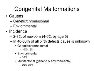
Causes: Inheritance: A multifactorial etiology has been assumed in which interaction between hereditary predisposition and environmental influences resultsin the defects.
Maternal factors - Maternal diabetes - Maternal phenylketonuria. - Maternal alcohol consumption and fetal alcohol syndrome - Genetic risk factors.
- A family history of a cardiac or noncardiac defect in either a parent or a preceding sibling is a major risk factor - Familial congenital heart defects are often concordant by phenotype and developmental mechanism
Genotype-phenotype correlation: - Careful family history of first- and second-degree relatives, including detailed analysis of pregnancy loss, racial origin, and consanguinity - A search for risk factors such as gestational diabetes mellitus
Murmurless Congenital Heart Diseases a) Cyanotic diseases - - Transposition of great arteries - Pulmonary atresia with intact septum - total anomalies of pulmonary venous drainage - Tricuspid atresia with or without pulmonary atresia
b) Acyanotic diseases - Core triatriatum - Severe coarctation - Coronary artery originating from pulmonary Artery. - Endocardial fibroelastosis
c) Others -Single ventricle without obstruction. - Hypoplastic left heart syndrome
* Duct dependent lesions - These are lesions presenting very early in life that are dependent on ductal patency for survival These lesions fall into two main categories
A) Cyanotic lesions in which pulmonary blood flow is almost or totally dependent on the duct • Severe tetrallogy of fallot- - Pulmonary atresia with intact septum pulmonary atresia with VSD- Critical pulmonary stenosis-
B)Acyanotic lesions Aortic atresia. - Critical aortic stenosis- Iterrupted aortic arch - - Severe pre-or juxtaductal Coarctation-
The radiographic classification of CHD relies on: • Clinical information (cyanosis) 2- Plain film information (increased pulmonary vascularity, decreased pulmonary vascularity (
I ) Acyanotic CHD with increased pulmonary vascularity. Common denominator of these lesions is that there is a L-R shunt where pulmonary flow is greater than true aortic flow;
The shunt can belocated in: 1- Ventricular septal defect. 2- Atrial septal defect. 3- Patent ductus arteriosus
4- Uncommon lesions: - Aorticopulmonary window. -Ruptured sinus of Valsalva aneurysm, coronary artery fistula 5-Other - Endocardial cushion defect - PAPVC
II) Acyanotic CHD with normal pulmonary vascularity Normal pulmonary vascularity is associated with either outflow obstruction or valvular insufficiency:
A)Outflow obstruction: • -Coarctation of aorta • - Interruption of aortic arch • - Aortic stenosis • - Pulmonary stenosis / insufficiency B) Valvular insufficiency: • - Aortic insufficiency • - Pulmonary insufficiency
III) Cyanotic CHD with decreased pulmonary vascularity - Common denominator of these lesions is that there is a decreased pulmonary vascularity due to obstruction of pulmonary blood flow. -In addition, there is an intracardiac defect through which blood is shunted away from the lungs causing cyanosis.
* Types: Normal heart size - Tetralogy of Fallot - Fallot variants ( trilogy, pulmonary atresia / pseudotruncus I/II)
Increased heart size -Ebstein's anomaly (largest heart in CHD) - Tricuspid atresia ddx: 1 - Uhl's disease (RV myocardium absent with tricuspid atresia) 2-Pulmonary stenosis with ASD
IV) Cyanotic CHD with increased pulmonary vascularity (admixture lesions) -Common denominator of these lesions is that there is an "admixture" of systemic and pulmonary venous blood (bidirectional shunting. -
-Clinical symtoms: CHF, cyanosis, recurrent pneumonia, growth retardation. -The admixture of venous blood may occur at the level of:
* Large veins: - Total anomalous pulmonary venous connection (ASD is also present) * Large arteries: - Truncus arteriosus
Atrium - Transposition of great arteries (VSD is also present). Ventricle - Single ventricle - DORV (types I, II = Taussig-Bing.
TETRALOGY VARIANTS 1- Pink tetralogy: -1/3have mild pulmonary valvular obstruction with large VSD, allowing sufficient pulmonary flow. - Pulmonary atresia and VSD = pseudotruncus, extreme end of the spectrum. - Infundibular hypertrophy in VSD (3%)
2- Pentalogy of Fallot: Tetralogy + ASD 3- Trilogy of Fallot: PA stenosis + RV hypertrophy +Patent foramen ovale , or (ASD)
TRANSPOSITION OF GREAT ARTERIES (TGA) Types: Complete transposition of great arteries (D-TGA) AORTA ORIGINATES FROM RV - PA originates from LV - Normal position of atria and ventricles
Corrected transposition of great arteries (L-TGA): -Transposition of great arteries - Inversion of ventricles - The relative position of aorta and pulmonary artery can be derived from the diagram on the right.
D-TGA (COMPLETE TRANSPOSITION): Two independent circulations exist: • Blood returning from body RV blood delivered to body (aberrant aorta) • Blood returning from lung LV blood delivered to lung (via ASD,etc).
This circulatory pattern is incompatible with life unless there are associated anomalies that permit mixing of the two circulations, e.g., through ASD, VSD, or PDA. •Hemodynamics: depends on the level of admixture and R->L shunting. •RA and RV enlarged.
GENERAL: Situs stuff: - Abdominal situs refers to position of liver and stomach: a) Abdominal situs solitus: liver on right, stomach on left (normal) b) Abdominal situs inversus: liver on left, stomach on right
c) Thoracic situs refers to position of the tracheobronchial tree:
Thoracic situs solitus (normal) 1-Left main bronchus longer than right main bronchus 2-Left main bronchus inferior to left PA 3-Right main bronchus superior to right PA
Type I( situs inversus totalis)with 1- Mirror-like malposition of the heart 2 - Mirror-like malposition of other viscera. 3 - Kartegner suyndrome: Situs inversus , sinusitis & bronciectasis
Type II:( Isolated dextrocardia) 1- Mirror-like cardiac malposition. 1- Normal position of other viscera. 3- serious cardiac anomalies.
TypeIII ( Dextroversion with): 1- Heart is merely displaced to right. 2- RV remains to right & LV remains to left. 3- Serious cardiac anomalies.
Type IV (Dextroposition) : Aquired dextrocardia: The heart is displaced by external factors: Pulmonary , pleural or diaphragmatic
The three common cynotic Heart Diseases 1) Falot tetralogy 2) fallot Trilogy. 3) Eisenmenger”s syndrome