Download
1 / 34
340 likes | 497 Views
Post-treatment surveillance in patients with esophageal cancer: Is it beneficial and worth the cost?. Moderators: David Cort, MD Alex Denes, MD Panelists: Stephen Swisher, MD, PhD Edward Lin, MD. Staging of Esophageal Cancer. Tumor staging T1- confined to mucosa/submucosa

E N D
Post-treatment surveillance in patients with esophageal cancer: Is it beneficial and worth the cost? Moderators: David Cort, MD Alex Denes, MD Panelists: Stephen Swisher, MD, PhD Edward Lin, MD
Staging of Esophageal Cancer Tumor staging T1- confined to mucosa/submucosa T2- extends to muscularis propria T3- extends into surrounding tissue T4 - involves major vessels, pleura, pericardium Nodal staging N0 - no nodes involved N1 - local nodes involved N2 - distant nodes involved
Treatment modalities for esophageal cancer • Esophagectomy • Combined modality • Pre-operative chemo-radiation >>> esophagectomy • Esophagectomy >>>> Post-operative chemo-radiation • Non surgical • Definitive chemoradiation
Results of surgical Resection for Esophageal Cancer Surgical marginsPathology margins • R0 No tumor No tumor • R1 No tumor Microscopic tumor present • R2 Tumor present Macroscopic tumor present
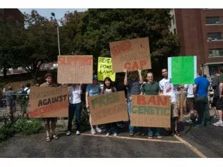
Post treatment follow up in esophageal cancer Ed Lin- 7 mins • What are the usual sites of recurrence • Local • distant • Benefits • Palliative chemo ± radiation • survival benefit • Quality of life • Treatment of recurrence in lymph node outside the initial field of initial radiotherapy • How- • Physical Exam- what signs to look for • CT chest/abdomen- what findings to look for • EGD – what symptoms should prompt it • Serum CEA levels- ? In which patients • EUS - ? role • How often • Suggested protocols for follow up
Case 1 • A 52 year old accountant with known history of Barrett’s esophagus and symptomatic reflux • surveillance endoscopy. • 5 cm segment of Barrett’s esophagus proximal to the GE junction. • Biopsy - Multiple foci of HGD • 1.5 cm sessile polypoid lesion at the GEJ • Biopsy- Invasive adenocarcinoma.
T1- confined to the mucosa and submucosa and sparing the muscularis propria. • N0 – no enlarged lymph nodes
Case 1 (continued) • PET/CT: • No nodal or distant metastases • He undergoes esophagectomy without complications • (R0 resection) • Surg path: • T1, N0, (M0) moderately differentiated adenocarcinoma, • No lympho-vascular infiltration • multiple foci of Barrett’s • all margins clear of tumor
AQ1. Appropriate post treatment follow-up of this patient would involve • CT chest and abdomen every 3 months • CT chest and abdomen every 6 months • CXR every 3 months • EGD every 3 months • All of the above • None of the above
Questions to panel • What are the chances of tumor recurrence • What are the usual sites of recurrence • Local • Treatment options • Benefits • Distant • Treatment options • Benefits • Suggested follow up after treatment
Ed Lin T1N0 GEJ • The cure rate 80-90%. • If EMR or radiation cure rate 60-70% (then regular EGD is indicated). • Q 6 months for the first 2 years, then annual physical exams with routine blood work. • Imaging only when clinically indicated.
Stephen Swisher • Repeat endoscopy 1 year after surgery to rule out residual Barrett’s or dysplasia • No CT Scan, CXR or PET scan unless symptoms because of low likelihood of distant mets with T1N0, LVI negative
Stephen Swisher • Repeat endoscopy 1 year after surgery to rule out residual Barrett’s or dysplasia • No CT Scan, CXR or PET scan unless symptoms because of low likelihood of distant mets with T1N0, LVI negative
A 65 year old house-wife with history of GERD presents with progressive dysphagia • EGD: • An irregular, non obstructing, ulcerated mass in the distal esophagus. • Biopsy: Moderately differentiated adenocarcinoma • EUS: • T3 tumor (infiltrating muscularis propria) • No enlarged lymph nodes • PET/CT: • intense FDG uptake in the distal esophageal mass • no lymph node or distant metastases
Case 2 ( continued) • Planned treatment: • Pre-operative chemoradiation followed by surgery • Patient recieves • combined modality therapy with radiation and chemotherapy • Follow up EGD: • no residual mass and biopsy shows only radiation effect. Patient is now reluctant about proceeding with esophagectomy
ARS Q2. Appropriate next step in this patient • Convince the patient to proceed with surgery as originally planned • Give additional chemo-radiation to full dose • Can wait and see how the patient performs • None of the above
CRT > RT; RT = S; CMT > S?; • RTOG-Hersovic: Chemo-RT > RT: 5-year 32%, vs 12% 20% vs 0% 10 year survival. LR > 45% • Intergroup 0123: 50.4 Gy > 64.8 Gy • Phase III: Modern RT = S • CMT vs S: OR 0.53-0.86. Three Meta-analysis (Ref 1-3). (Many small studies isolated positive study mostly with 5FU/Cisplatin/50.4 cGy. Urschel Am J Surgery 2003:6:553. 1. Surgery 2005; 137:1727 2. Gut 2004;7:925 3. Walsh et la. NEJM.1997 Kelsen DP NEJM 1998; Yu ASCO 2006 Abst 4012
Questions to Panel • Chances of tumor recurrence • Sites of tumor recurrence • Local • distant • Treatment options • Salvage esophagectomy • Suggested follow up
Stephen Swisher • CT or CT/PET and endoscopy with biopsies q 3 months x2 then 6month x 3 then yearly (RTOG 0246) • Early follow/up similar survival to trimodality
4 weeks later, patient changes his mind and wants to proceed with the planned esophagectomy • Esophagectomy • R0 resection • Path: • No residual carcinoma in the esophagus • 12 lymph nodes are clear
Questions to panel • What are the chances of tumor recurrence • What are the usual sites of recurrence • Local • Treatment options • Benefits • Distant • Treatment options • Benefits • Suggested follow up after treatment
Case 3 • A 48 year old high school teacher presents with progressive dysphagia and weight loss • EGD: • large ulcerated nearly circuferential mass in the lower third of the esophagus. • Biopsy: Moderate to poorly differentiated adenocarcinoma with lymphovascular infiltration. • PET/CT: • Intense FDG uptake in paraesophageal lymph nodes. • No distant metastases. • EUS: • T3 tumor (Nearly circumferential mass, extension into the adventitia) • N1 (multiple enlarged regional lymph nodes)
Case 3 Endoscopic appearance T3N1 tumor
Case 3 (continued) • Treatment: • combined modality therapy with radiation and chemotherapy. • Follow up EGD • 75% regression of the mass. • Biopsy: residual adenocarcinoma. • Esophagectomy • R0 resection • Path: • Residual moderately differentiated adenocarcinoma, • foci of carcinoma in 3 regional lymph nodes.
AQ. 3 What is the best management option now? • Follow up with EGD and CT scan every 3 months • Follow up with EGD and CT scan every 6 months • Additional radiation therapy to maximal dose • Combination salvage chemoXRT
Questions to panel • What are the chances of tumor recurrence • What are the usual sites of recurrence • Local • Treatment options • Benefits • Distant • Treatment options • Benefits • Suggested follow up after treatment
Case 4 • A 68 year old retired carpenter with a history of CAD, CABG, CHF, COPD, and DM presents with progressive GERD symptoms. • No dysphagia or weight loss. • EGD: • distal esophagitis with an area of ulceration just proximal to the GE junction • Biopsy: Moderately differentiated adenocarcinoma. • EUS: • T2 N0tumor
Case 4 ( continued) • PET/CT: • No abnormal FDG uptake in paraesophageal lymph nodes. • No distant metastases. • Surgical evaluation: • Not candidate for resection due to co-morbidities • Treatment: • Completes full course of combined chemotherapy and radiation.
Questions to panel • What are the chances of tumor recurrence • What are the usual sites of recurrence: 40% • Local • Treatment options: • Benefits • Distant • Treatment options • Benefits • Suggested follow up after treatment