Download
1 / 64
640 likes | 874 Views
Innate Host Defenses Immunology - I. April 10, 2007. Adaptive defenses. Antigens are produced in response to the presence of non self or foreign antigens. Innate Defenses. The defense mechanisms that are active against any invading agent

E N D
Innate Host DefensesImmunology - I April 10, 2007
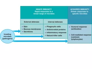
Adaptive defenses • Antigens are produced in response to the presence of non self or foreign antigens
Innate Defenses • The defense mechanisms that are active against any invading agent • These are often prior to the activation of adaptive defenses
Physical Barriers • Skin – External covering. Layered to prevent invasion. NO vascularity in the epidermis. Outer layer keratinized • Mucous membranes- the presence of mucous prevents invasion in the respiratory tree.( These are produced by cells in the pseudostratified epithelial layer) Cilia and hairs assist in the removal of organisms from these surfaces • Physical removal results from coughing • Flushing can occur via vomiting and diarrhea
Chemical Barriers I • Sweat is high in salt and electrolyte content. This prohibits the growth of many microbes • Sebum is produced by sebaceous glands and results in the elimination of some bacteria from the skin due to the acidity of the secretion
Chemical Barriers • Acidity of stomach destroys many microbes that are sensitive to acidic pH • Lysozyme is present in tears and causes the lysis of bacterial cells • Lactoferrin and transferrin prevent the binding of Fe by bacteria that have high requirements for the ion
Cellular Defenses I • Granulocytes • Basophils – release histamine that helps in initiating the inflammatory response • Mast cells which also release histamines • Eosinophils that may detoxify foreign substances
Eosinophils • Eosinophils-- 10 to 15 um diameter • Eosinophils constitute 2.0 to 4.0% of leukocytes. These cells usually contain a bilobate (two lobes) nucleus and a cytoplasm full of brightly stained eosinophilic (orange-red) specific granules. • Eosinophils function specifically as phagocytes to destroy larvae of parasites that have invaded tissues i.e. in trichinosis, schistosomiasis, and appear to play a role in allergic responses. • Other functions of eosinophils include phagocytosis of antigen antibody complexes.
Neutrophils • Size—12–15 µ in diameter approximatetly 2.5 times the diameter of a RBC. • Nucleus—lobulated or partially segmented with dark purple, dense chromatin. • Cytoplasm—smooth, pale pink or light blue, finely granular. • 50-70% of circulating WBC
Cellular Defenses II • Neutrophils – Polymorphonuclear leukocytes that guard the skin and the mucous membranes. These are active, respond quickly, and phagocytic. • Dendritic cells are cells with long membrane extensions that are also phagocytic
Basophils • Basophils are the least common of the white blood cells. • By their large cytoplasmic granules which obscure the nucleus in stained preparations as shown above. • They have many similarities with mast cells and actually become mast cells on leaving the blood and entering surrounding tissues. • Mast cells are widely distributed throughout the body and are commonly found in close proximity to the walls of small blood vessels. • Both basophils and mast cells have highly specific receptors for IgE produced in response to various allergens.
Phagocytes • Monocytes migrate from the bone marrow into the blood • When these cells move from the blood into the tissues they undergo a series of cellular changes and become macrophages. These cells are capable of destroying cellular debris as well as individual cells by phagocytosis
Steps in Phagocytosis • Recognition of invading microorganisms • These recognize molecular patterns unique to the pathogen such as petidoglycan or flagellin proteins • Macrophages and dendritic cells are can recognize Gram Positive and Gram Negative Bacteria
Chemokines • Cytokines are chemicals that have specific roles in host defenses including the cells involved in the inflammatory response • Chemokines are a class of cytokines that attract phagocytes to the site of an infection • A strategy for avoiding ingestion is to interfere with phagocytosis
Adherence • Avoidance of phagocytosis – the formation of an antiphagocytic capsule • This is evident in Streptococcus pneumonia ( classic example of pahtogenicity) • The immune system seaks to prevent his by caoting the microbe with antibodies or with proteins of the complement system
Ingestion • Once captured, the phagocytes ingest the microbe y producing fingerlike extensions that surround the microbe • The microbe is incorporated into a vesicle called the phagosome In this way it is walled off from the cell’ cytoplasm
Digestion • The phagosome can then merge with a lysosome of the host cell • This forms a phagolysosome • The pH of the interior is acidic when the enzymes that are located in the lysosome are dumped into the phagosome • Defensins are a category of molcules that are present in the lsyosomes that perforates the outer membrane
Destruction of microbes • The production of oxidative radicals such as hydrogen peroxide and superoxide ions are also produced • These radicals destroy the cell membrane
Residual bodies • The remnants of bacterial cells that are indigestible are left over in the cytoplasm • The remnants can remain in the residual body
Yersinia pestis • The causative agent of plague forms a capsule that are not vulnerable to destruction by macrophages • The capsule prevents digestion by the enzymes and molecules in the lysosome
Mycobacterium • The engulfed bacterium resides in a membrane enclosed fluid-filled compartment called a parsiophorous vacuole( PV) that do not fuse with a lysosome. • Lysosomal enzymes cannot unable to digest the PVs
Toxins • Microbes also produce leukocidins that are released and destroy WBC • Streptolysins are molecules that are released by streptococci to injure WBC
Extracellular killing • Larger parasites are killed by products secreted by defensin cells • Eosinphils secrete a major basic protein that is capable of interacting with the worms outer covering so that the parasite is destroyed and the phagocytes can ingest particles that are left over
Defensins • There are three types of defensins produced by mammalian cells • Alpha, Beta, and Theta • Help to maintain balance of natural microflora in the body • Found on body surfaces – exterior and interior
Natural Killer Cells • Natural Killer Cells respond to viral infections • Their activity is accelerated by the presence of cytokines • They recognize glycoproteins on the outside of infected cells • Dumb their contents into the infected cells
Lymphatic System • Network of vessels, nodes, and tissues that are closely associated with the cardiovascular system • It collects excess fluid from the spaces between the body cells • It transports digested fats to the cardiovascular system • It supports innate and adaptive immune responses
Lymphatic circulation • Lymphatic capillaries are found throughout the body • They are larger in idamter than blood capillaries • They collect the excell fluid and plasma proteins that leak between the cells • The fluid inside of the capillaries is referred to as lymph
Lymph nodes • Fluid circulates through the lymphatic capillaries to the lymphatic vessels • It then passes through the lymph nodes. • It is returned to the venous blood through he right and left lymphatic ducts which drain the fluids into the right and the left subclavian veins
Route of lymph flow though a lymph node • Afferent lymphatic vessel • Subscapular sinus • Trabecular sinus • Medullary sinus • Efferent lymphatic vessel
Cells that are found in the Lymph Node • Cells in the Medulla- Plasma cells, B cells, and Macrophages • Cells of Inner Cortex – T cells and Dendritic Cells • Cells in Germinal Center – B cells, Macrophages
Thymus Gland • Multilobed lymphatic organ located beneath the sternum • The thymus is very active at birth and processes lymphocytes and release them into the blood as T cells
Spleen • Upper left quadrant • Spleen is similar in structure to lymph nodes • Contains B and T cells as well as erythrocytes
GALT • Gut associated lymphoid tissue • Major sites of antigen production against microbes that are mucosal pathogens
Inflammation • Inflammatory processes are directed against the invaders • They cause changes in tissue • Help to kill and eliminate the invaders • Assist in the repair of tissues
Histamine • Histamnes are released by basophils and mast cells when tissues are damaged • Histamine causes vasodilation( the diameter of the blood vessels increase) • The membranes become more permeable
Vasodilation • Increases the blood flow to the damaged area • It causes the area infected or affected to become warm and red • Fluids may accumulate around the injured cells causing swelling( edema)
Tumor Necrosis Factor ( alpha) • In conjunction with histamine release • The fluid that enters the injured tissue carries the chemical components of the blood clotting mechanism • If the injury has caused bleeding platelets and clotting factors such as fibrin are present
Bradykinin • Peptide that stimulates the pain at the injured site • Prostaglandins may also be released in this process