Download
1 / 23
240 likes | 463 Views
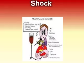
Block 17: Cardiopulmonary resuscitation, shock management, transportation and alcohol in trauma . A Engelbrecht 2013. Three measures makes a big difference:. High quality CPR Early defibrillation Treating reversible causes. Cardiopulmonary resuscitation.

E N D
Block 17: Cardiopulmonary resuscitation, shock management, transportation and alcohol in trauma A Engelbrecht 2013
Three measures makes a big difference: • High quality CPR • Early defibrillation • Treating reversible causes
Cardiopulmonary resuscitation • Most victims of out of hospital sudden cardiac arrest do not receive any bystander CPR • Why do you think this is the case? • What can be done to improve on this? • Continued emphasis on high quality CPR • Compression rate of at least 100/min (a change from “approximately” 100/min • Compression depth of at least 2 inches (5cm) in adults and children and 1.5inches (4cm) in infants
Change from A-B-C to C-A-B • Who is excluded from this sequence? • The vast majority of adult cardiac arrest cases are? • Who has the highest survival rate from cardiac arrest? • What are the critical elements of resuscitation in these cases? • CPR are delayed by responders retrieving barrier devices and assembling ventilation equipment
Chain of survival • Should you call for help first or first do one cycle of chest compressions?
Chain of survival includes: • Recognition of cardiac arrest • Activation of the EMS • Early CPR • Rapid Defibrillation • Effective advanced life support • Integrated post cardiac arrest care
What to do if a bystander is not trained in CPR? • Hands only CPR • Compressions only CPR • “Push hard and Fast” • Continue until AED arrives or EMS arrives and take over care of the victim • If trained to provide rescue breaths – 30 compressions to 2 breaths • Survival rates from cardiac arrest of cardiac aetiology are similar for hands only and compressions plus rescue breaths
Major chances in advanced cardiovascular life support • Quantitative wave form capnography is recommended for monitoring of endotracheal tube placement and CPR quality • Atropine is no longer recommended for routine use in the management of PEA
End tidal CO2 and termination of resus • End tidal CO2 monitoring confirm the futility of resuscitation and forecast likelihood of resuscitation • End-tidal carbon dioxide level of 10 mmHg or less measured 20 minutes after the initiation of advanced cardiac life support accurately predicts death in patients with cardiac arrest associated with electrical activity but no pulse • Cardiopulmonary resuscitation may reasonably be terminated in such patients – • Ref: Levine R, End-tidal Carbon Dioxide and Outcome of Out-of-Hospital Cardiac Arrest, New England Journal of Medicine.
Medications in ALS • Atropine is no longer reccomended for routine use in PEA or asystole and has been removed from the ACLS cardiac arrest algorithm • When would atropine be appropriate for use in cardiac arrest?
Organised post cardiac arrest care: • New section in the 2010 AHA guidelines • Comprehensive structures system • Cardiopulmonary and neurological support • Theraputic hypothermia • PCI • Tapering inspired oxygen concentration after ROSC based on saturation • Maintain saturation > 94% • Avoid hyperoxia • At SO2 of 100% the PO2 can vary between 80 and 500
Important causes for cardiac arrest in Trauma • Hypoxia – airway obstruction - ↓ LOC; breathing problems – tension pneumothorax flail chest etc. • Hypoperfusion – cardiac tamponade / hypovolaemia • Primary rhythm is usually PEA or asystole. VF not as common
Management • Focus pre-hospital resuscitation to safely extricate and attempt to stabilize patient and minimize interventions that delay transport to definitive care \ • On scene: blunt trauma and cardiac arrest. Poor prognosis. Stop CPR if no response in 5 minutes • Penetrating trauma – better prognosis • Primary survey C-A-B-C management
Emergency Department Thoracotomies: • 2002 report: resuscitative thoracotomies for trauma patients in the ED, the 3 survivors of 10 victims of penetrating trauma all had signs of life and vital signs on arrival at the ED. In contrast, all 19 patients with blunt trauma died, despite the fact that 14 of the 19 “had vital signs” at the time of the thoracotomy • In a database of 959 resuscitative thoracotomies, 22 victims of penetrating trauma and 4 victims of blunt trauma survived to hospital discharge after receiving prehospital CPR (overall survival rate of 3%). • In 2001 the Committee on Trauma of the American College of Surgeons published a systematic review of 42 studies of ED thoracotomies involving nearly 7000 patients, published from 1966 to 1999. In this database, survival was 11% (500 of 4482) for victims of penetrating trauma and 1.6% (35 of 2193) for victims of blunt trauma.
Suggested Indications for Resuscitative Thoracotomy: Patients With Traumatic Cardiac Arrest
Damage control resuscitation • Permissive hypotension • Haemostatic resuscitation • Damage control surgery
Transportation of the trauma patient • Reasons for transporting a patient – Inter- or intrahospital transport. • Need for more advanced diagnostic procedures e.g. CT, MRI • Need for more advanced therapeutic procedures e.g. surgery • Transfers to a tertiary facility
Potential complications/risks associated with transportation • Equipment failure/ malfunction • Dislodgment of tubes such as thoracostomy tube, Foley catheter, surgical drain • Disconnection from ECG or ventilator • Power failure • Failure of suction apparatus • Respiratory system • Hypo- or hyperventilation • Acute desaturation & drop in PaO2 • Acid base disturbances • Airway loss/problem • Cardiovascular system • Hypotension • Hemorrhage • Arrhythmias • Loss of vascular access • Neurological deterioration • Increase in intracranial pressure • Decrease in cerebral perfusion • Hypoxia • Other: • Death e.g. acute pulmonary embolus • Collision risk • Temperature changes
Planning/preparation • Earlier transport rather than later as soon as the decision is made to transport the patient. • The patient must be stable for transportation: resuscitation before transport (with crystalloids or blood products as indicated). • Correct possible electrolyte disturbances & precipitants. • Inspect all invasive catheters and establish vascular access as necessary. Remove air from all intravenous catheters and bags. • Inspect the airway for position and stability. Restabilize or reposition as necessary. • Review radiographs and laboratory data. • When necessary, perform procedures such as bronchoscopy, central catheter insertion, or thoracostomy tube insertion before departure.2 • Minimize continuous medication infusions to necessary vasoactives and volume expanders.2 • Discontinue infusions of paralytics and/or sedatives; those agents are given by bolus during transport.2 • Do not move the patient until the blood gases are considered satisfactory and hemodynamic stability has been achieved or optimized.
Communication: • Direct communication between teams on both sides: physician-to-physician and nurse-to-nurse.2 • Make sure the area where the patient is meant to be moved is ready to receive him/her.1 • Adequate documentation – incl. transport permission forms and test results must accompany the patient. • Driver/ pilot must be aware of all the hospitals located between the sending and receiving hospitals.2
Personnel • One of the professionals should be the nurse in charge of the patient, with experience in CPR or specially trained in transport of patients in serious conditions. • In accordance with the serious condition and instability of the patient, the second professional can be a doctor, nurse or paramedic. • A doctor should accompany the patient who presents with physiological instability and who might need an urgent action.
Modes of transport • Choice depends on transport distance, medical condition of the patient, staff and resources availability, weather forecast and necessary medical procedures during transport.1 • In case of air transport it is also important to be aware of possible physiological changes regarding the altitude and its influence on clinical features. • 0 - 200 km: road ambulance or helicopter if urgent. • 200 - 300 km: road ambulance, helicopter or fixed-wing air ambulance depending on condition. • > 300 km: fixed-wing air ambulance. • Scheduled (commercial) service for stable patients only.
Alcohol in trauma • Role of alcohol • Alcohol Hampers Diagnosis • Other complications: electrolyte and fluid imbalance, blood coagulation problems, cardiomyopathy, hepatic dysfunction, and alcohol withdrawal • Bias against treating an inebriated patient who is uncooperative and disruptive may lead to quick disposition of that case in order to free up time for more "deserving" patients