Download
1 / 36
360 likes | 448 Views
Have Microscopes Become Obsolete in Our Universities. Dr Camile S Farah. Microscope vs Microscopy. The difference between the microscope and microscopy is largely the difference between a technology and an academic discipline.

E N D
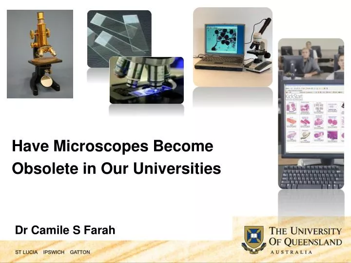
Have Microscopes Become Obsolete in Our Universities Dr Camile S Farah
Microscope vs Microscopy • The difference between the microscope and microscopy is largely the difference between a technology and an academic discipline. • A microscope is the material entity that sits on our laboratory and lecture benches while microscopy is that set of concepts that marshal this material technology into a means of viewing, debating and understanding the microscopic world beyond human vision.
Microscopy of pathological specimens is a staple of practical classes used to teach pathology and illuminate disease processes. • The pre-eminence of the light microscope as a teaching device has only recently been challenged by medical and dental academics.
This change has been necessitated by: • financial restrictions • a reduction in the amount of time allocated to instruction in histology & pathology • a major shift in the design of new curricula to include newer learning approaches such as problem based learning (PBL) and constructivist approaches • and “Baby Boomer” staff members retiring, with fewer “Generation X” colleagues taking up academic posts
Crucially, students generally dislike using light microscopes for the examination of tissue specimens. • They find using the light microscope difficult, frustrating, and tiresome. • Even when students are shown how to use the microscope, they complain about the quality and consistency of its use. • Consequently students are notengaging deeply in course content where the light microscope is present and only do the bare minimum to pass the course; while some are unable to do that.
Proper use of a light microscope and its accompanying glass slide is not that technically challenging, but it does require practice and a helping hand from a knowledgeable tutor. • Furthermore, light microscopes need qualified technical staff to both maintain them, and the numerous glass slide sets that are required for practical classes. Traditionally this has required a large amount of space and technical assistance.
Introducing virtual microscopy (VM) into medical, dental and veterinary programs offers a remedy to these problems while at the same time presenting both technical and pedagogic challenges of its own. • Here I will highlight our experience with the introduction of virtual microscopy and computer-assisted student-centered learning in histology & pathology in our dental program.
What is Virtual Microscopy • With this new technology, glass microscopic slides are scanned at high magnification (x40) and viewed on a computer window browser as virtual slides, and stored on DVDs or accessed over the Internet. • The virtual images are completely maneuverable in any direction, and at any magnification up to ×40. • In essence the computer becomes the microscope.
+ =
ImageScope (Free Viewer) • Image Viewing • Zoom in • Pan • Slide Annotations • Digital Slide Conferencing • Take high-resolution snapshots for publications
WebScope/Webviewer Web-based Cross-platform (PC and Mac) Slide annotations No installation on local machine
Vital Statistics • VM has been implemented in the dental program at UQ since 2005 • 2nd, 3rd and 4th year courses that teach histology and pathology now use it • We have accumulated data over a 4 year period from a total of 393 respondents using a LIKART scale (data presented as % in agreement)
Vital Statistics • Qualitative feedback was also accumulated, and focus groups undertaken • VM images were delivered first on DVD then on a dedicated school server in a designated computer lab. Now they are available over the Internet using KickStart software. • Blackboard (Information Management System) is used to deliver autopsy cases, and to communicate with students • Autopsy cases are streamlined with the VM images, so both programs are run side by side
Results • Results from the end of course surveys regarding the use of virtual microscopy showed that 71.43% of students agreed that ‘the virtual microscope positively enhanced their learning of the course material’.
Results • Another pertinent statistic revolves around the question of VM helping students to better ‘understand the course material during scheduled laboratory sessions’. 77.19% of students agreed with this statement.
Results • 91.07% of students felt that ‘having access to the autopsy cases on Blackboard and access to the virtual microscope slides together enhancedtheir learning of the material in the course’.
Results • Given that VM technology is both spatially and temporally variable in its delivery of microscopic content, it is interesting to note that 92.85% of students agreed that the virtual microscope helped them ‘understand the course material outside of laboratory sessions’.
Results • The students’ preference for the virtual microscope is explained by its effective software(92.98%), ease of navigation (70.18%), resolution (71.93%), and magnification (78.95%), and its interface with the autopsy manual on Blackboard (71.43%). • In addition, up to 90% of students thought that using the virtual microscope was FUN.
Results • 59.65% of students felt that using the virtual microscopepositivelyaffected their grade for the course, in contrast to only 22.81% stating that the light microscope positively affected their grade. • The students’ subjective feelings regarding the positive effect of the virtual microscope on their grades was paralleled in part by the objective measures of their performance assessment tasks.
Results • When asked if ‘using the virtual microscope positively enhanced my learning of the material in this course’, up to 88.34% agreed with this core statement. • These are significantly convincing figures that indicate the extent of just how much more deeply engaged students are in studying microscopy via digital means.
Results • At the heart of the question of whether or not microscopes have become obsolete in our universities is the most potent statement students were asked to respond to: ‘virtual microscopy technology should be expanded to eliminate completely the need for light microscopy in teaching microscopy’, only 42.11% agreed. • Although students clearly expressed a preference for VM, this was not at the insistence of the complete elimination of light microscopy.
Discussion Points • One thing that cannot be easily disputed in the current pedagogical transformations going on in the tertiary sector is the intensely symbiotic relationship between traditional educational ideas centred on didactic teaching and learning (which generally have come under the rubric of ‘literacy’), and electronic media (which now has ‘electracy’ as a term to define its educational parameters).
Discussion Points • It was Marshall McLuhan who wrote ‘that the content of any medium is always another medium’. • The transition from the analogue technology of the light microscope to digital technology of VM exemplifies this idea in practice: the analogue imagery of the light microscope forms the very basis through which VM develops and proceeds.
Discussion Points • Even if in the longer term, light microscopy does become obsolete as a physical technology, the ideas and concepts it helped propagate most certainly will not disappear. • It is this evolutionary change (as opposed to change that is a revolutionary either/or choice) that sits at the heart of student unease about the statement: ‘The virtual microscopy technology should be expanded to eliminate completely the need for light microscopy’.
Discussion Points • While there certainly are, and will be, difficulties with the introduction of VM technology into a curriculum, our data shows that it has been a positive move in the UQ context. • Although file sizes are large, the software implements a compression algorithm to facilitate downloading of the images over the Internet. This is evidenced by the fact that most students (91.23%) felt that the download speeds of the virtual microscopy material were acceptable.
Discussion Points • It remains to be tested though whether we as educators are prepared not only for the technological challenges but also the conceptual changes wrought by this new technology • It is certainly not business as usual when the computer becomes the new microscope as well as the means via which a redefinition of microscopy as a discipline might proceed.
Conclusions • We can say with confidence that students prefer VM to light microscopy. • The data shows that the virtual microscope positively altered how the students learnt and interacted with the course material, but this did not significantly affect what they learnt.
Conclusions • Although the students were able to learn the course content regardless of the visualizing modality used, the enhancement of the learning process by using VM speaks highly of its effectiveness in helping students engage and interact with the course material.
Conclusions • It is easy to become overwhelmed by the extent and the rate of technological change in higher education. This hectic pace of change, though, sometimes obscures broader conceptual reconfigurations going on in education and more broadly in society and culture.
Conclusions • The shift from the light microscope, whose tenure is far from over, to the virtual microscope signals these larger changes from print based forms of knowledge to electronic forms, and from teacher centred, or didactic delivery of course content, to more student centred frames of knowledge transfer.
Conclusions • It may not be immediately obvious to educators in the first instance but there can be little doubt that any technological innovation brings with it a change in the way we might think about a particular problem or issue.
Conclusions • It is especially important to grapple with these issues when ultimately it is through the health of our students’ patients that this transition from light microscopy to virtual microscopy will be tested.
The Plug! The story highlighted in this presentation originated from a dire situation where microscopy in the dental curriculum was dying a slow and agonizing death. The untenable situation required thorough review,and the need to revise the status quo. It lead to a refreshed look at the way we were doing things, and resulted in a renewal of microscopy, and of student engagement and excitement about learning.
Acknowledgements • Dr Terry Maybury – Project Officer ‘The Virtual Slidebox’. • Dr Maybury’s position as Project Officer is funded by an Australian Learning and Teaching Council (formerly Carrick Institute) grant awarded to CSF at the University of Queensland entitled ‘The Virtual Slidebox — A New Paradigm for Exploring the Microscopic World’ (CG7-467).